
- Title
-
Zebrafish Rfx4 controls dorsal and ventral midline formation in the neural tube
- Authors
- Sedykh, I., Keller, A.N., Yoon, B., Roberson, L., Moskvin, O.V., Grinblat, Y.
- Source
- Full text @ Dev. Dyn.

Rfx4 expression and mutagenesis. Representative zebrafish embryos stained using WISH to visualize distribution of rfx4 RNA show expression in the caudal neural tube, including the midbrain, hindbrain, and spinal cord at 5 s (A), 10 s (B), 14 s (C), and 22 s (D). Expression in the preoptic diencephalon is detectable by 10 s and strong by 22 s. There is no detectable expression in the Kupffer's vesicle (KV; asterisk in A). fb, forebrain; mb, midbrain; hb, hindbrain; sc, spinal cord. Arrowheads mark expression in preoptic diencephalon. E: CRISPR assays were used to target a site in the 8th exon of 18 rfx4 exons. Two mutant alleles of rfx4 were generated: uw8013, which encodes a 13‐nt deletion, and uw8110, which encodes a 10‐insertion, at the CRISPR target site. Location of the translation‐blocking morpholino (TBMO) is indicated by a line over the translation start site in exon 1. F: Schematic representation of the wildtype (full‐length) and mutant (truncated) rfx4 proteins. The conserved DNA binding winged‐helix domain (DBD; amino acids 61–136) is indicated in yellow; the conserved dimerization domain (DIM; amino acids 315–487) is shown in purple. The predicted proteins encoded by rfx4uw8013 and rfx4uw8110 are truncated due to frameshifts after amino acid 262; both truncated proteins lack the dimerization domain. EXPRESSION / LABELING:
|

Aberrant curvature in rfx4 mutant and morphant embryos. Heterozygous rfx4uw8013 embryos develop normally (A), while their homozygous siblings show pronounced downward curvature (B) by 2 dpf. C: PCR genotyping of individual progeny of rfx4uw8013/ + parents shows that the curved phenotype is linked to rfx4uw8013 homozygosity. D,E: Embryos injected with translation‐blocking morpholino targeting rfx4 develop with downward curvature. F: The aberrant curved morphology in rfx4 morphants is independent of apoptosis. Summarized from four experiments. e, embryos. |
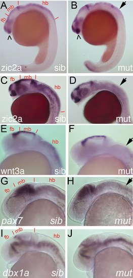
Dorsal hindbrain is aberrant in rfx4 mutant embryos by 24 hpf. WISH was used to visualize gene expression patterns in rfx4uw8013 homozygotes vs rfx4uw8013/ + sibling embryos, which develop normally, at 24 hpf. zic2a expression is reduced in the mutant dorsal hindbrain by 18 s (A,B) and remains depleted at 24 hpf (C,D). E,F: wnt3a expression is unaffected in midbrain, but reduced in hindbrain of mutant embryos. G,H: pax7a expression is unaffected in the alar plate of the midbrain and hindbrain in mutant embryos. I,J: dbx1a expression in the diencephalon and in the midbrain/hindbrain basal plate is likewise unaffected in mutants. mb, midbrain; hb, hindbrain; open arrowheads, optic stalk expression; arrows, depleted hindbrain expression. |
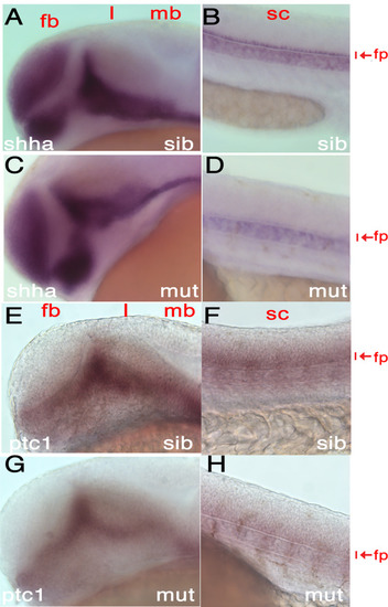
Ventral floor plate is aberrant in rfx4 mutant embryos by 24 hpf. In rfx4uw8013/+ sibling embryos, shha is expressed in the cranial primordium (A) and in the spinal cord floor plate, adjacent to notochord (B). Cranial expression of shha is unchanged in rfx4uw8013 homozygotes (C), while its expression in the spinal cord floor plate is strongly reduced (D). E,F: Expression pattern of ptc1, a target of Shh signaling, resembles that of shha in normal siblings. G,H: ptc1 expression is reduced in mutant spinal cord. fp, floor plate; sc, spinal cord. |
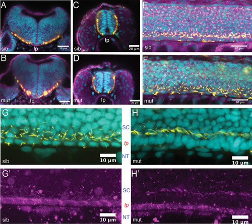
Ciliogenesis is not overtly affected in the spinal cord of rfx4 mutant embryos. A–F: 24‐hpf embryos derived from an rfx4uw8013/+ incross were immunostained for acetylated tubulin (yellow) and gamma‐tubulin (purple) to visualize ciliary axonemes and basal bodies, respectively. Nuclei were counterstained with DAPI (cyan). Confocal cross‐sections through the hindbrain (A,B) and spinal cord (C,D). E: confocal sagittal z‐stack through the spinal cord of a heterozygous sibling shows normal distribution of primary (short) and motile (long) cilia. F: Equivalent z‐stack through a homozygous mutant sibling shows presence of both primary and motile cilia. G,H′: rfx4uw8013/+ incross progeny were immunostained for acetylated tubulin (yellow in G,H) and a motile cilia component rsph9 (purple; G′, H′) to visualize ciliary axonemes of primary vs motile cilia. Nuclei were counterstained with DAPI (cyan). G: confocal sagittal z‐stack through the ventral portion of the spinal cord of a heterozygous sibling shows normal medial floor plate morphology and normal distribution of motile (long) cilia produced by both medial and lateral floorplate. H: Equivalent z‐stack through a homozygous mutant sibling shows presence of long cilia in the absence of identifiable floor plate. fp, floor plate; SC, spinal cord; NT, notochord. Scale bar = 30 μm in A,B; 20 μm in C–F; 10 μm in G,H. PHENOTYPE:
|

RNAseq transcriptome analysis identifies a set of Rfx4‐dependent targets in 24‐hpf zebrafish. A: Embryos derived from rfx4uw8013/+ parents were sorted by morphology into mutant and sibling groups. RNA extracted from individual embryos (4 normal and 5 curved) was used to prepare cDNA libraries for illumina high‐throughput sequencing. B: The heat‐map colors represent relative expression levels of genes identified as differentially expressed, i.e., genes assigned value of False‐Discovery Rate below 0.05. C–L: Embryos derived from rfx4uw8013/ + incrosses were stained at 1 dpf by WISH to visualize expression of foxa2 in the floor plate (C–H), atoh1a in the dorsal hindbrain (I,J) or wnt1 in the dorsal hindbrain and midbrain (K,L). C,D: foxa2 expression is disrupted in the ZLI (arrowhead) and floor plate (arrow) in mutant embryos. E,F: foxa2 expression is reduced in the medial and lateral floor plate in the mutant hindbrain (arrows). G,H: foxa2 expression is reduced in the spinal cord floor plate in mutant embryos. I,J: atoh1a expression is reduced in the mutant dorsal hindbrain. K,L: wnt1 expression is reduced in the mutant dorsal midbrain and hindbrain. Representative rfx48013 and rfx48013/+ embryos are shown dorsally, anterior to the left, except for C, D, G, H shown laterally. fb, forebrain; mb, midbrain; hb, hindbrain; sc, spinal cord; fp, floor plate; SC, spinal cord; NT, notochord; asterisk, mid‐hindbrain boundary; ZLI, zona limitans intrathalamica. |