
- Title
-
Accurate quantification of homologous recombination in zebrafish: brca2 deficiency as a paradigm
- Authors
- Vierstraete, J., Willaert, A., Vermassen, P., Coucke, P.J., Vral, A., Claes, K.B.M.
- Source
- Full text @ Sci. Rep.
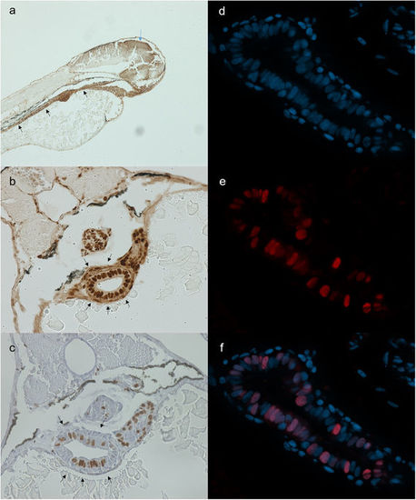
Stainings for PCNA (a,b), BrdU (c) and geminin (d-f). All stainings were performed on 72 hpf embryos. (a) Sagittal section stained for PCNA. Strong staining is found in the tectum (blue arrow) and gastro-intestinal tract (black arrows). (b) Transversal section stained for PCNA. PCNA stains positive in the gut tube, swim bladder and liver. (c) Transversal section stained for BrdU. BrdU intake is prominent in intestinal cells. (d–f) Separate images of nuclear staining (DAPI) (d), mCherry (zGem signal) (e) and overlay (f). (f) mCherry signal is present in a large proportion of intestinal cells. EXPRESSION / LABELING:
|

Representative images of Rad51 staining. Rad51 foci are absent in unirradiated sample (a), while irradiated sample displays clear Rad51 foci (b) (green fluorescence). (c) Schematic drawing of an unirradiated (left) and irradiated (right) gut tube section. Cells in blue are in late S-/G2-phase and thus capable of HR. Cells in white are not capable of HR and will never show Rad51 foci. Red dots indicate Rad51 foci. Quantifying all cells would lead to a serious underestimation of your signal, due to the large proportion of non-HR capable cells. Instead, quantifying only cells containing foci will give a more true value for irradiated conditions. To estimate the amount of HR capable cells in unirradiated samples, one can extrapolate the percentage of cells that contain Rad51 foci from the irradiated samples. EXPRESSION / LABELING:
PHENOTYPE:
|

Rad51/geminin co-staining of irradiated embryo. Rad51 foci (green) are almost exclusively found in geminin positive nuclei (red). EXPRESSION / LABELING:
PHENOTYPE:
|
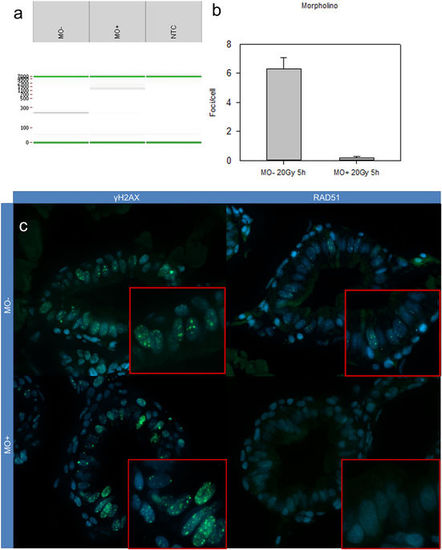
Results of morpholino experiment. (a) Digital gel of RT-PCR (obtained by Labchip®GX software) of uninjected (MO−) and injected (MO+) embryos. Injecting the morpholino results in retention of intron 7, resulting in a transcript containing a premature stop codon. This is visualised as a longer RT-PCR fragment, using primers located in exon 4 and 8 (expected size: 250 bp; in case of intron 7 retention: 1600 bp). In the morphants, there is a clear band of 1600bp, indicative for intron retention. (b) Quantification of Rad51 foci in three morphants (MO+) and uninjected (MO−) embryos. Almost no Rad51 foci are present in the morphants. Error bars display 95% CI. (c) γH2AX foci are present in both irradiated wild types (upper left) and morphants (lower left). Wild types display clear Rad51 foci (upper right), while morphants display no Rad51 foci. EXPRESSION / LABELING:
PHENOTYPE:
|

ZFIN is incorporating published figure images and captions as part of an ongoing project. Figures from some publications have not yet been curated, or are not available for display because of copyright restrictions. EXPRESSION / LABELING:
PHENOTYPE:
|
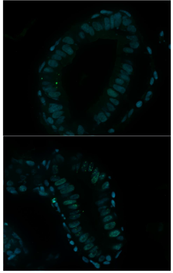
γH2AX staining. Unirradiated (upper) and irradiated (lower) gut section of 72 hpf embryo. Dose administered was 20 Gy, followed by fixation at 5 hpi. |