
- Title
-
Patient-derived xenograft in zebrafish embryos: a new platform for translational research in gastric cancer
- Authors
- Wu, J.Q., Zhai, J., Li, C.Y., Tan, A.M., Wei, P., Shen, L.Z., He, M.F.
- Source
- Full text @ J. Exp. Clin. Cancer Res.

Gastric cancer cells survived and induced angiogenesis in larval zebrafish (fli-eGFP). a Typical images of subintestinal vessels of uninjected embryo at 3 dpf. AGS cells (b) and SGC-7901 cells (c) were injected to the zebrafish embryos, and induced angiogenesis at 1 dpi. 50 nM VRI can block angiogenesis of the subintestinal vessels caused by cell lines AGS (d) and SGC-7901 (e). The white boxes at lower right corner showed the higher magnification of the upper left white boxes. The arrow indicated the tumor cell-induced angiogenesis. Hpf: hours post fertilization; dpi: days post injection EXPRESSION / LABELING:
|
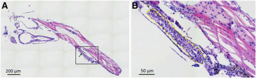
Histological hematoxylin and eosin (H & E) staining of the whole-mount zPDX model at 7 dpi. A low (a) and a higher (b) magnification of a representative zPDX were showed. Black box in (A) indicates the area of zoom. Arrows in (A) and yellow dashed line in (B) point to the adenoid structure formed by primary epithelial cells from GC patient. Dpi: days post injection |

Primary cells from GC tissue induced angiogenesis and metastasized in larval zebrafish (fli-eGFP). 600–800 primary cells for patient samples were fluorescently labeled in red and microinjected into yolk sac of each zebrafish embryo at 48 hpf (0 dpi). Primary cells from GC tissue induced angiogenesis at 1 dpi (a), and showed invasive behaviors at 1 dpi (b), 4 dpi (c) and 7 dpi (d) in zebrafish PDX model. At 7 dpi, we could detect cancer cells in the brain (e), and the caudal hematopoietic tissue (CHT) region (f). Arrow in (a) points to the tumor induced new blood vessels within tumor mass. Arrows in (c)-(f) point to the metastasized tumor cells in the head, trunk and tail of the zebrafish embryos. Hpf: hours post fertilization, dpi: days post injection EXPRESSION / LABELING:
|
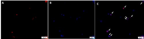
Fluorescent microscopy analysis of dissociated embryos. Xenografted embryos were dissociated and the resulting cell suspension were analyzed by fluorescent microscopy. The eight cells in the field of view that stain positive for CM-DiI colocalize with individual nuclei (white arrows) stained with DRAQ5 nuclear stain. |