
- Title
-
Developmental and adult characterization of secretagogin expressing amacrine cells in zebrafish retina
- Authors
- Dudczig, S., Currie, P.D., Jusuf, P.R.
- Source
- Full text @ PLoS One
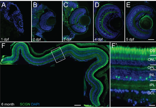
Secretagogin expression in embryonic and adult zebrafish retina. Micrographs of vertical sections through zebrafish retina immunohistochemically labeled for secretagogin (SCGN–green) with nuclei counterstained by DAPI (blue). (A–E) Sections through retinas at 1–5 days post fertilization (dpf) show earliest secretagogin positive cells detected at 3 dpf (C) and maintained at subsequent days. (F) Collage through retinal section in 6 month old zebrafish. Secretagogin expression in the amacrine layer in the inner half of the inner nuclear layer (INL) remains strong throughout adulthood. (F') Higher magnification inset of boxed region in F shows secretagogin labeled with stained processes showing monostratified band in the center of the inner plexiform layer (IPL). OS: outer segments; ONL: outer nuclear layer; OPL: outer plexiform layer; GCL: ganglion cell layer. Scale bar (E) for A-E is 50 μm, scale bar (F) is 100 μm, scale bar (F') is 20 μm. |
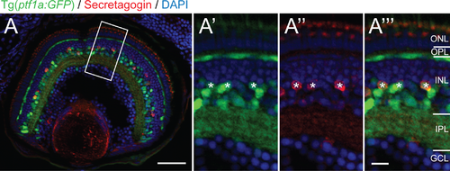
Secretagogin positive cells co-label with the Ptf1a:GFP amacrine marker in the inner nuclear layer. Micrograph at 5 days postfertilization showing secretagogin immunostained Tg(ptf1a:GFP) zebrafish retinas. Higher magnification of boxed view of boxed inset shows co-localization (asterisks) of SCGN+ (red) and Ptf1a:GFP+ (green) within individual cells marked in the green (A'), red (A'') and double (A''') channels.). ONL: outer nuclear layer; OPL: outer plexiform layer; INL: inner nuclear layer; IPL: inner plexiform layer; GCL: ganglion cell layer. Scale bar (A) is 50 μm, scale bar (A''') for A'–A''' is 10 μm. |

Secretagogin positive cells from a regular mosaic with highest density along the horizontal midline. (A, B) Micrograph collage and schematic showing secretagogin immunostaining in the inner nuclear layer of a flat mounted whole adult zebrafish retina. (C, D) Density of secretagogin labeled cells across the nasal-temporal (F) or dorsal-ventral (G) axes indicate high even density along the horizontal meridian, with the density along the dorsal-ventral axis peaking in central retina and decreasing towards the periphery (n = 20–21 ROIs for each of n = 3 adult eyes). Density was counted in ROIs (200 μm x 200 μm) every 250 μm until the edge of the retina. (E–G) Individual examples showing nearest neighbor analysis of region of interest (200 μm x 200 μm) indicated by boxed regions in B. Secretagogin labeled cells at any eccentricity are distributed regularly. ROIs were located at 250 μm, 750 μm and 1250 μm distance from the optic nerve center. Scale bar (A) is 200 μm. |

Comparative expression of secretagogin and other calcium binding proteins within the zebrafish retina. (A–C) Micrographs showing cross-sections through zebrafish retina at 5 days postfertilization. Higher magnification of boxed regions in each row show secretagogin expression in green and other calcium binding protein expression in red: Parvalbumin (PV–A), Calbindin (CB–B), Calretinin (CR–C). (D–F) Pie charts show quantification of singe and double labeling (asterisks). Secretagogin labeled cells are mutually exclusive from parvalbumin (D) expressing cells, but overlap partially with calbindin (E) and represent a subpopulation of calretinin (F) expressing cells. Scale bar (C) for A–C is 50 μm, scale bar (C''') for A'–C''' is 20 μm. |

Secretagogin amacrine subtype markers and other calcium binding proteins within the zebrafish retina. (A–F) Micrographs showing cross-sections through zebrafish retina at 5 days postfertilization. Insets show separate red and green channels of subregion of the double channel images. Co-labeling (asterisks) of secretagogin—SCGN (green) with amacrine subtypes markers (red): GABA (A), tyrosine hydroxylase–TH (B), serotonin– 5HT (C), choline acetyltransferase–ChAT (D), neuropeptide Y—NY (E), Sox2 (F). Most SCGN+ cells show little co-localization with other markers (white dots). (G) Graph shows proportion of secretagogin cells that also co-label for the other amacrine markers (asterisks). Scale bar (D) for A–F is 20 μm. |