
- Title
-
Rapid functional analysis of computationally complex rare human IRF6 gene variants using a novel zebrafish model
- Authors
- Li, E.B., Truong, D., Hallett, S.A., Mukherjee, K., Schutte, B.C., Liao, E.C.
- Source
- Full text @ PLoS Genet.
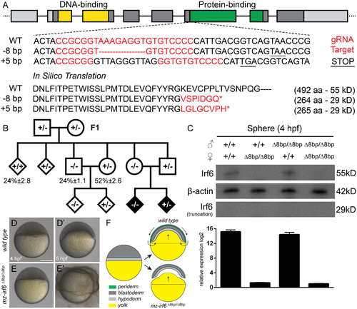
Generation and characterization of the zebrafish irf6 CRISPR mutant model. (A) Zebrafish irf6 gene structure composed of eight exons, a helix-turn-helix DNA-binding domain (yellow), and a SMIR/IAD protein-binding domain (green). The CRISPR gRNA target site was located in exon 6 at the start of the protein-binding domain. Sanger sequencing of the target site revealed a -8bp deletion (Δ8bp) that created a frameshift and premature stop codon, truncating the protein to 29 kD as predicted by in silico translation. Another +5bp insertion was also identified. (B) Breeding pedigree revealed the irf6 mutant phenotype in F3 and the importance of maternal transcripts. (C) Top: western blot at the sphere stage (4 hpf) revealed a lack of Irf6 full-length (55 kD) or truncated (29 kD) protein in all maternal irf6 Δ8bp/Δ8bp embryos but not paternal irf6 Δ8bp/Δ8bp embryos or wild type embryos. Bottom: relative gene expression by RT-qPCR revealed a lack of irf6 mRNA transcripts in all maternal irf6 Δ8bp/Δ8bp embryos but comparable levels between wild type and paternal irf6 Δ8bp/Δ8bp embryos. Error bar = 2xSEM, n = 3. (D-D’) Wild type embryos at the sphere stage (4 hpf) (D) and at the 30% epiboly stage (5 hpf) (D’). (E-E’) Maternal irf6 Δ8bp/Δ8bp embryos at the sphere stage (4 hpf) (E) and displaying the periderm rupture phenotype at 5 hpf (E’). Scale bar = 250 μm. (F) Cross-sectional schematic through the embryonic midline illustrating the zebrafish embryo epiboly process. Arrows represent cell and yolk directional movements. Wild type embryos experience rapid cellular lamination and yolk doming between 4–5 hpf, while maternal-zygotic irf6 Δ8bp/Δ8bp embryos experience incomplete periderm differentiation and animal pole/yolk separation. EXPRESSION / LABELING:
PHENOTYPE:
|
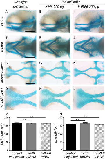
Periderm rupture and craniofacial development can be rescued by injection of either zebrafish or human wild type IRF6 mRNA. (A-L) Zebrafish embryos at 96 hpf stained with alcian blue for cartilaginous craniofacial elements. Maternal/zygotic-null irf6 -/- embryos were rescued by microinjection of either zebrafish irf6 mRNA (E-H) or human IRF6 mRNA (I-L) at the one-cell stage, preventing the periderm rupture phenotype and restoring normal craniofacial development compared to wild type embryos (A-D). Scale bar = 150 μm. (M-N) Dimensional measurements of dissected ethmoid plates at 96 hpf, with length (l) and width (w) denoted by dashed lines on panel (D). The length (M) and width (N) of ethmoid plates from zebrafish and human IRF6 mRNA rescued maternal/zygotic-null irf6 -/- embryos are statistically indistinguishable in dimensions compared those of wild type embryos. Error bar = 2xSEM, n = 12. |

Maternal-null irf6-/- embryo gene expression is rescued by injection of either zebrafish or human wild type IRF6 mRNA. (A-H) Whole-mount in situ hybridization analysis of wild type embryos (A-D) compared to maternal/zygotic-null irf6 -/- embryos (E-H) at the sphere stage revealed strong down-regulation of critical irf6 downstream genes such as krt4 and grhl3. Scale bar = 150 μm. (I) Relative gene expression of wild type embryos, maternal/zygotic-null irf6 -/- embryos, and maternal/zygotic-null irf6 -/- embryos rescued with either wild type zebrafish or human IRF6 mRNA (100 pg) microinjections, for a panel of genes with crucial roles in the irf6 gene regulatory network. Error bar = 2xSEM, n = 3. |
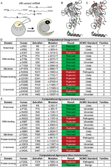
Functional characterization of human IRF6 missense gene variant protein functions with the zebrafish irf6 model. (A) Experimental approach for characterizing protein functions of human IRF6 missense gene variants. Variant mRNAs were synthesized and microinjected into maternal-null irf6 -/- embryos at the one-cell stage and assessed for phenotypic rescue at 24 hpf. (B-C) Protein modeling of the protein-binding domain and C-terminus of IRF6 using ExPASy with crystalline structures of IRF1. (B) is mapped with missense variant amino acid residues (green) whose mRNA rescued the periderm rupture phenotype, while (C) is mapped with missense variant amino acid residues (red) whose mRNA failed to rescue. (D-F) Results for functional rescue of periderm rupture with maternal-null irf6 -/- embryos for representative human IRF6 missense gene variants. Results were classified as rescued if any maternal-null irf6 -/- embryos injected with variant mRNA remained alive and phenotypically wild type at 24 hpf (50 embryos/round, n = 3). Missense gene variants were categorized by location within the IRF6 protein, and by computational results from PolyPhen-2 and SIFT on whether the in silico predictions agreed on the deleterious effects of the missense gene variants on protein function. Further shown are ACMG guideline pathogenicity predictions (pathogenic, likely pathogenic, uncertain, and benign), and the number of families identified for each variant (all gene variant annotations were based on NM_006147.3). |
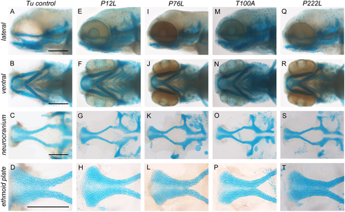
IRF6 missense gene variants can rescue periderm rupture and restore normal craniofacial development. (A-T) Craniofacial morphologies of maternal-null irf6 -/- embryos rescued by human IRF6 missense gene variant mRNA microinjections (100 pg/embryo) at 96 hpf stained with alcian blue. (A-D) Uninjected wild type control. (E-H) p.P12L. (I-L) p.P76L. (M-P) p.T100A. (Q-T) p.P222L. Scale bars = 150 μm, n = 3. |