
- Title
-
Histological and transcriptomic effects of 17α-methyltestosterone on zebrafish gonad development
- Authors
- Lee, S.L.J., Horsfield, J.A., Black, M.A., Rutherford, K., Fisher, A., Gemmell, N.J.
- Source
- Full text @ BMC Genomics

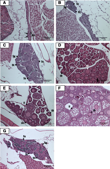
Histological analysis of gonads of 40 dpf control (a and b) and MT-treated juvenile zebrafish (c-f). a Presumptive testis characterised by the presence of residual bodies (RB), an indicator of oocyte degeneration, infiltration of stromal (Str) cells and spermatogenic tubule-like structures (→). b Ovary with Stage IA (IA) and Stage IB (IB) oocytes. c Transforming gonad. d Immature testis with spermatogonia (Sg), spermatocytes (Sc) and spermatids (St). e Mature testis possessing spermatogonia (Sg), spermatocytes (Sc), spermatids (St) and spermatozoa (Sz). f Ovary with Stage IA (IA) and Stage IB (IB) oocytes. Bar = 100 μm |
Histological analysis of gonads of 60 dpf untreated control (a - f) and MT-treated juvenile zebrafish (g). a Presumptive testis comprising infiltration of stromal (Str) cells and spermatogenic tubule-like structures (→). b Immature testis containing spermatogonia (Sg), spermatocytes (Sc) and spermatids (St). c Mature testis containing spermatogonia (Sg), spermatocytes (Sc), spermatids (St) and spermatozoa (Sz). d Ovary with Stage IA (IA) and Stage IB (IB) oocytes. e Ovary with Stage I IA (IA), Stage IB (IB) and Stage II (II) oocytes. f Ovary with Stage I IA (IA), Stage IB (IB) and Stage II (II) oocytes. g Mature testis possessing spermatogonia (Sg), spermatocytes (Sc), spermatids (St) and spermatozoa (Sz). Bar = 100 μm |