
- Title
-
nr3c1 null mutant zebrafish are viable and reveal DNA-binding-independent activities of the glucocorticoid receptor
- Authors
- Facchinello, N., Skobo, T., Meneghetti, G., Colletti, E., Dinarello, A., Tiso, N., Costa, R., Gioacchini, G., Carnevali, O., Argenton, F., Colombo, L., Dalla Valle, L.
- Source
- Full text @ Sci. Rep.

ZFIN is incorporating published figure images and captions as part of an ongoing project. Figures from some publications have not yet been curated, or are not available for display because of copyright restrictions. PHENOTYPE:
|
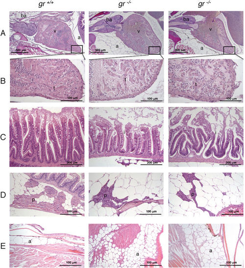
Histology of 8-month-old gr −/− and gr +/+ zebrafish. All histological images of 8-month-old gr −/− and gr +/+ zebrafish were taken from longitudinal sections stained with haematoxylin and eosin (H&E). The left panels represent wild type fish, whereas the middle and right panels show tissues from two mutant fish. (A) heart; (B) particular of the heart at higher magnification showing reduced trabecular network in the mutant samples; (C) intestinal mucosa with sloughing epithelium at the villous tips and reduced height of villi in mutants; (D) visceral view showing reduced extension of pancreas in mutants; (E) consistent increase of subcutaneous adipose tissue in mutants. ba = bulbus arteriosus; v = ventricle; a = adipose tissue; i = intestine; p = pancreas; t = trabeculae. PHENOTYPE:
|

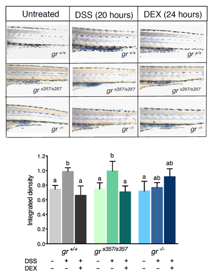
gr s357/s357 and gr −/− 5-dpf zebrafish larvae in transgenic Tg(9xGCRE-HSV.Ul23:EGFP) background cannot respond to DEX treatment. (A) Fluorescence microscopy images of gr +/+, gr s357/s357 and gr −/− 5-dpf zebrafish larvae in transgenic Tg(9xGCRE-HSV.Ul23:EGFP)ia20 background. Treated larvae were subjected to 10 μM DEX treatment for 24 h (from 4 dpf to 5 dpf). (B) Integrated density analysis of fluorescence of 5-dpf zebrafish larvae of the three genotypes with or without DEX treatment. Homozygous zebrafish mutants deriving from heterozygous or homozygous parents were analysed separately. Values represent the mean ± SEM. Asterisks indicate that expression levels are significantly different from the control (two-way-ANOVA, *P < 0.05, ***P < 0.001). n = 15 larvae for each group. (C) qRT-PCR analysis of fkbp5, foxo3b and mmp-9 in gr +/+, gr s357/s357 and gr −/− 5-dpf zebrafish larvae deriving from homozygous parents with or without DEX treatment. Values represent the mean ± SEM. Different letters indicate statistically significant differences checked by two-way ANOVA followed by Tukey’s multiple-comparison test. fkbp5 (P < 0.01); foxo3b and mmp-9 (P < 0.001). Data were generated from four biological replicates. |
|
ZFIN is incorporating published figure images and captions as part of an ongoing project. Figures from some publications have not yet been curated, or are not available for display because of copyright restrictions. |
|
ZFIN is incorporating published figure images and captions as part of an ongoing project. Figures from some publications have not yet been curated, or are not available for display because of copyright restrictions. |
|
ZFIN is incorporating published figure images and captions as part of an ongoing project. Figures from some publications have not yet been curated, or are not available for display because of copyright restrictions. |

(A): qRT-PCR of gr mRNA in 5-dpf mutant larvae compared to control shows a statistically significant reduction of gr expression. Values represent the mean ± SEM. Asterisks indicate that expression levels are significantly different from the control: ***P < 0.001. Data were generated from four biological replicates. (B): Representative images of 5-dpf control gr+/+ and mutant gr-/- larvae after exposure to VBA stimulus. gr-/- mutants appear darker in comparison to control. (C): Representative gel image of PCR genotyping using genomic DNA from tail fins of adults born from a cross between gr heterozygotes. (D): Western blot of liver proteins from 8-month-old gr-/- and gr+/+ zebrafish showing disappearance of the protein band with respect to control. |

(A) Histological analysis of two samples of each genotype, gr+/+, grs357/s357 and gr-/- zebrafish, at 45 dpf of age. All histological images were taken from longitudinal sections stained with haematoxylin and eosin (H&E). Panels compare tissues and structures in the three different genotypes. Top 3 panels present a total body section of one sample of each genotype. Middle 9 panels present details of the gonads, head kidneys and pharyngeal teeth. Bottom 3 panels show a longitudinal section of the heart showing the reduced trabecular network of the gr-/- heart ventricle. |

(B) Histological analysis of two samples of each genotype, gr+/+, grs357/s357 and gr-/- zebrafish, at 45 dpf of age. All histological images were taken from longitudinal sections stained with haematoxylin and eosin (H&E). Top 3 panels present sections of esophageal sacs and proximal intestine. Middle 6 panels present details of the intestine showing the presence of a thinner epithelium in gr-/- samples. Bottom 6 panels show details of the endocrine and exocrine pancreas. |

(C) Histological analysis of two samples of each genotype, gr+/+, grs357/s357 and gr-/- zebrafish, at 45 dpf of age. All histological images were taken from longitudinal sections stained with haematoxylin and eosin (H&E). Top 3 panels present sections of spinal cord and vertebral column together with the kidney. Middle 3 panels present details of pseudobranch and gills. Bottom 3 panels show sections of the liver. |
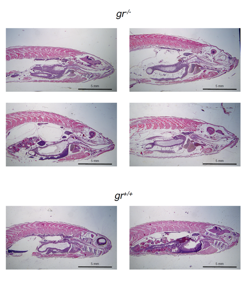
Longitudinal sections stained with haematoxylin and eosin (H&E) of four gr-/- and two gr+/+ to better visualize the adipose tissue increase in mutants. |
(A): Whole-mounts of the posterior intestinal region of control, DSSexposed and DSS plus DEX-exposed larvae of the three genotypes analysed after staining with alcian blue. (B): Comparison of alcian blue-stained mucous granules in the intestine of the above larvae. n = 15 larvae for each group. Values represent the mean ± SEM. Different letters indicate statistically significant differences checked by two-way ANOVA followed by Fisher’s post hoc test (p<0.05). |