
- Title
-
greb1 regulates convergent extension movement and pituitary development in zebrafish
- Authors
- Li, S.Z., Liu, W., Li, Z., Li, W.H., Wang, Y., Zhou, L., Gui, J.F.
- Source
- Full text @ Gene

Expression patterns of greb1 in tissues and embryos. (A, B) Real time PCR detection of greb1 transcripts in adult tissues (A) and embryos of different developmental stages (B). (C–L) Whole mount in situ hybridization of greb1 transcript in embryos at different development stages including (C) sphere, (D) 30% epiboly, (E) shield, (F) bud, (G) 3S (3-somite), (H) 10S (10-somite), (I) 21S (21-somite), (J) 24 hpf (prim-5), (K) 30 hpf (prim-15) and (L) 36 hpf (prim-23). The embryos are lateral views with animal pole toward the top and dorsal to the right. EXPRESSION / LABELING:
|

Embryonic phenotypes of WT (A, D, G, J), greb1−/− (B, E, H, K) and rescued (greb1−/− + RNA) (C, F, I, L) groups at the indicated stages. K1 and K2 respectively represent the severe and light development defect embryos at 26 hpf. |
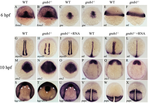
Whole mount in situ hybridization detection of different marker genes in WT, greb1−/− and rescued (greb1−/− + RNA) embryos at 6 hpf and 10 hpf. A and B, foxa3; C and D, gsc; E and F, ntl; G–I, myoD (staining the adaxial cell); J–L, ntl (staining the forerunner cell group, axial chorda mesoderm); M–O, otx2 (staining the anterior axial hypoblast and neural plate); P–R, zic1 (staining the neural plate); S–U, dlx3b (white arrowheads) and hgg (staining the prechordal plate, indicated by white arrows); V–X, papc (staining the paraxial mesoderm). Embryos in panels A–F were shown in lateral view with dorsal side on the right, and embryos in panels G–X were shown in dorsal views with the anterior to the top. |

Survival ratio (A), body length (B), body weight (C), and 1, 2 and 3 month old size (D) comparisons between WT and greb1−/− mutant. Scale bars: 5 mm. |

Whole mount in situ hybridization examination of different pituitary cell markers in WT, greb1−/− and rescued (greb1−/− + RNA) embryos. A, lim3; B, pit1; C, gh; D, tshβ; E, prl; F, gthα; G, pomca. The embryos are shown in lateral views with the dorsal to the right (A and B), and in dorsal views with the anterior to the top (B–G). |

ZFIN is incorporating published figure images and captions as part of an ongoing project. Figures from some publications have not yet been curated, or are not available for display because of copyright restrictions. |
|
ZFIN is incorporating published figure images and captions as part of an ongoing project. Figures from some publications have not yet been curated, or are not available for display because of copyright restrictions. |
Reprinted from Gene, 627, Li, S.Z., Liu, W., Li, Z., Li, W.H., Wang, Y., Zhou, L., Gui, J.F., greb1 regulates convergent extension movement and pituitary development in zebrafish, 176-187, Copyright (2017) with permission from Elsevier. Full text @ Gene