
- Title
-
Essential but partially redundant roles for POU4F1/Brn-3a and POU4F2/Brn-3b transcription factors in the developing heart
- Authors
- Maskell, L.J., Qamar, K., Babakr, A.A., Hawkins, T.A., Heads, R.J., Budhram-Mahadeo, V.S.
- Source
- Full text @ Cell Death Dis.
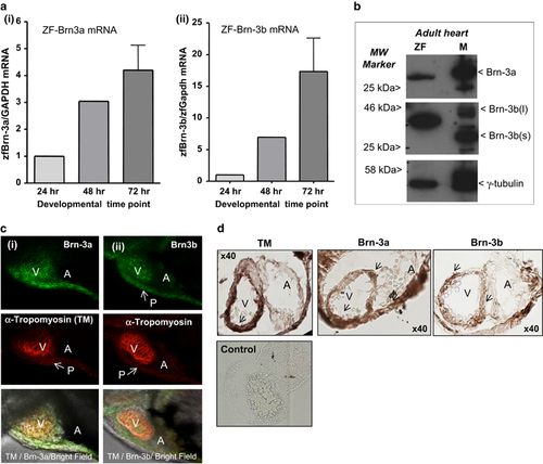
(a) Results of qRT-PCR showing (i) Brn-3a and (ii) Brn-3b mRNA levels in the developing zebrafish at 24, 48 and 72 hpf. cDNA from total RNA was amplified using primers to ZF Brn-3a and Brn-3b and a variation in mRNA levels was corrected using ZF GAPDH. Values were expressed as fold induction relative to expression at 24 h (set at 1). (b) Representative western blot analysis showing single protein band for both Brn-3a and Brn-3b in extracts from adult ZF compared with adult mouse heart (M), used as a positive control. MW markers indicate the protein size and gamma (γ) tubulin was used to control for variation in total protein. (c) Representative images showing whole-mount immunostaining for (i) Brn-3a or (ii) Brn-3b (green; top panels) in ZF hearts at 72 hpf. Co-staining with tropomyosin (red; middle panels) indicate cardiomyocytes in the developing heart. Lower panel shows merge with bright field image. V, A, P (indicated by arrow). (d) Representative images of DAB-immunostained ZF embryos sections at 72 hpf. Protein localisation is seen as dark brown staining in ventricles (indicated by arrow), identified by TM, also shows Brn-3a and Brn-3b expression. × 40 magnification. A, atria; hpf, hours post fertilisation; MW, molecular weight; P, pericardial sac; TM, tropomyosin; V, ventricle; ZF, zebrafish heart |
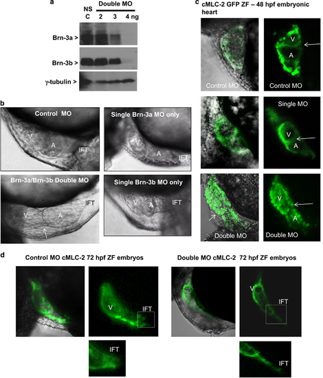
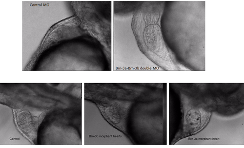
(a) Western blot analysis showing changes in Brn-3a or Brn-3b proteins extracts prepared from embryos at 72 hpf following injection with different amounts of oligonucleotide MO used to target both Brn-3a and Brn-3b, in fertilised eggs. Gamma (γ) tubulin was used to control for variation in total protein. (b) Photomicrographs showing bright field images of zebrafish heart at 48 hpf following injection of control non-silencing MO only, Brn-3a MO only, Brn-3b MO only or both Brn-3a and Brn-3b (double MO). The failure to loop, which results in linear double morphant heart is indicated by arrow. (c) Representative images showing results of similar studies carried out in CMLC2-GFP, in which the heart is marked with green fluorescent proteins. Left panels show merge of bright field and GFP, and right panels show GFP only in embryonic hearts taken from embryos following injection with control MO, single MO or double MO to target both Brn-3a and Brn-3b. (d) Representative images of hearts from ZF embryos at 72 hpf following injection with control MO or double MO is shown to highlight the significant changes in inflow tract when both Brn-3a and Brn-3b are targeted. A=atria; hpf, hours post fertilisation; IFT, inflow tract; MO, morpholinos; V, ventricle EXPRESSION / LABELING:
PHENOTYPE:
|
Video showing heart function in zebrafish embryos targeted with morpholino oligonucleotides to reduce Brn-3a and Brn-3b compared with non specific control or single morphants targeted for only Brn-3a or Brn-3b PHENOTYPE:
|