
- Title
-
Requirement for Jagged1-Notch2 signaling in patterning the bones of the mouse and human middle ear
- Authors
- Teng, C.S., Yen, H.Y., Barske, L., Smith, B., Llamas, J., Segil, N., Go, J., Sanchez-Lara, P.A., Maxson, R.E., Crump, J.G.
- Source
- Full text @ Sci. Rep.

Craniofacial and middle ear defects in mice deficient for Jag1 or Notch2. (a) Diagrams of the heads of zebrafish, mouse, and human (left to right) show homology between the fish jaw skeleton and the mammalian middle ear ossicles. The fish hyomandibula is homologous to the mammalian stapes (green); the fish palatoquadrate is homologous in part to the mammalian incus (red); and the proximal portion of the fish Meckel’s cartilage is homologous to the mammalian malleus (brown). (b) Micro-CT scans of mouse skulls at three weeks of age. Compared to control Notch2 f/f mice, Wnt1-Cre; Jag1 f/f and Wnt1-Cre; Notch2 f/f mice exhibit a persistent foramen (arrowheads in dorsal view) and midfacial hyperplasia resulting in an abnormally shaped skull and malocclusion (lateral view). (c,d) Dissected middle ear ossicles of three-week-old mice stained with Alizarin Red S. Compared to Jag1 f/+ controls, Wnt1-Cre; Jag1 f/f and Wnt1-Cre; Notch2 f/f mice display a fully penetrant columellar stapes phenotype. Wnt1-Cre; Notch2 f/f mice also rarely display an ectopic process from the anterior medial edge of the incus (arrow). Compared to wild-type siblings, some Jag1 +/− mice display a columellar stapes and a small ectopic process from the posterior medial edge of the incus body (arrow and inset). |
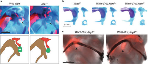
Mispatterning of middle ear cartilages and formation of the stapedial artery in Jag1-deficient mice. (a,b) Newborn mice were stained with Alcian Blue for cartilage and Alizarin Red S for bone. Close-up views show the developing middle ear, which is diagrammed below for wild-type and Jag1 heterozygous mice (malleus, brown; incus, red; stapes, green). Dissected middle ear cartilages are shown for conditional mutants. Arrows point to the stapes cartilage, which is reduced in size in both heterozygous and conditional Jag1 mutant mice. (c) The stapes of Wnt1-Cre; Jag1 f/+; Rosa26-Tomato and Wnt1-Cre; Jag1 f/f; Rosa26-Tomato mice fluoresce red and the stapedial arteries appear black from India ink injection. The artery is still present in Jag1-CKO mice, where it deviates around the misshapen stapes cartilage. Arrowheads point to the stapes. |
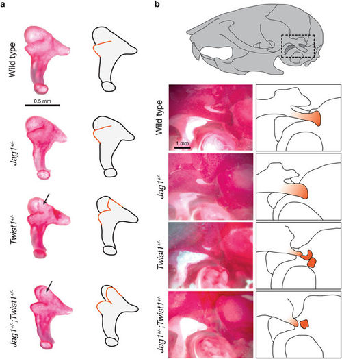
Incus and retrotympanic defects in Jag1; Twist1 compound mutants. (a) Dissected incus bones of three-week-old mice stained with Alizarin Red S. Wild types and this Jag1 +/− example display a normal incus. In contrast, Twist1 +/− and Jag1 +/−; Twist1 +/− mice have an extra process (black arrows) extending from the anterior medial edge of the incus body. Accompanying diagrams illustrate the ectopic processes with orange lines. (b) Views of the temporal bone in three-week-old mice stained with Alizarin Red S. The dashed box in the illustration shows the approximate region being imaged. Compared to wild-type and Jag1 +/− mice, Twist1 +/− and Jag1 +/−; Twist1 +/− mice display reduction and fragmentation of the retrotympanic process (shown in orange in the adjacent diagrams). |

Hearing loss and middle ear defects in patients heterozygous for JAG1 mutations and/or diagnosed with AGS. (a) The ratio of types of hearing loss for the right and left ears were calculated based on the 44 subjects tested. The degrees of hearing loss encompassed in each type of hearing loss were also analyzed. Table S1 lists the degrees of hearing loss as a range indicating the loss at its best frequency and worst frequency. The categorical analyses illustrated by the pie charts have taken into account only the loss at the worst frequency. (b) CT scans of the temporal bone in the axial plane from a control 69-year-old male without AGS and subject 5 who is heterozygous for a JAG1 loss-of-function mutation. Magnified areas of the dashed box regions and accompanying diagrams are shown below. Compared to the right stapes (orange) from the control subject, the right and left stapes of subject 5 appear as a single rod (i.e. columellar). Adjacent sections showed relatively normal articulation of the stapes with the incus. For better comparison, the orientation of the left stapes is flipped horizontally in the magnified view. (c) Audiogram of subject 5 (see Table 2) indicates mild to profound mixed hearing loss in the left ear and normal to mild sensorineural hearing loss with a potential high-frequency conductive component in the right ear. (d) Compared to a control 69-year-old male without AGS, CT scans of the temporal bone in the coronal plane show inappropriate ossification (red) of the oval window in the left ear of subject 1. The control right ear is flipped horizontally in the magnified area and accompanying diagram. |