
- Title
-
Live imaging of primary ocular vasculature formation in zebrafish
- Authors
- Hashiura, T., Kimura, E., Fujisawa, S., Oikawa, S., Nonaka, S., Kurosaka, D., Hitomi, J.
- Source
- Full text @ PLoS One

The primary ocular vasculature in zebrafish. (A–E) Ocular vascular anatomy of Tg(flk1:EGFP)k7 embryos at 2 dpf. Lateral (A, C, D, and E) and ventral (B) views. Lateral image of A divided into three parts: the superficial ocular (C) and hyaloid (D) vasculatures and the CVP (E). Panels C, D, and E in B indicate the projected region of each image. Arrows in B and D indicate the vascular plexus of the HA. Arrowheads in C indicate the IOC forming, which will connect the superficial ocular vasculature with the hyaloid vasculature. |

Arterial and venous cerebral angioblast clusters in zebrafish. Whole-mount in situ hybridization of WT embryos at 9S (C, D, I, and J), 12S (A, B, E, F, K, and L), 18S (G and H), 21S (M and N), and 26hpf (O). The expression of etsrp/etv2 (A and B), hey2 (C–H) and flt4 (I–O) genes was analyzed in both lateral view (A, C, E, G, I, K, M and O) and dorsal view of the cranial region (B, D, F, H, J, L and N). The differentiation of the aCAC and vCAC from the ROC and MOC was demonstrated. PCV: posterior cardinal vein. CV: caudal vein. |

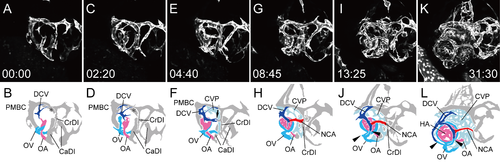
Primary ocular vasculature formation (dorsal view). Selected time-lapse images of Tg(flk1:EGFP)k7 embryo from 18S (S1 Movie) (A, C, E, G, I, and K) and their schematic diagrams (B, D, F, H, J, and L). The time (hours:minutes) from the first frame is labeled in each image (A, C, E, G, I, and K). Rostral is downward and the right side faces left. The formation of the left ocular vasculature was mainly observed. The formation of the hyaloid and ciliary vasculatures was demonstrated. Ocular vessels in the schematic diagrams are colored (NAC: red, DCV: blue, OA: pink, and OV: sky blue). Arrowheads in D indicate the rostral branch from the vCAC (PMBC), which anastomosed with the cranial division (CrDI) from the aCAC. Arrow in F indicate the medial branch from the vCAC (PMBC), which extended dorsally and anastomosed with the opposite branch at the midline to form the ACeV. |

The OV from the vCAC formed the vascular plexus surrounding the OA. Selected time-lapse images of a living Tg(flk1:EGFP)k7 embryo from 18S (S2 Movie) (A, C, E, G, and I) and their schematic diagrams (B, D, F, H, and J). The time (hours:minutes) from the first frame is labeled in each image (A, C, E, G, and I). Rostral is downward and the right side faces left. The formation of the left ocular vasculature was mainly observed. To visualize ventral vascular formation, only ventral slices were extracted from S1 Movie. Ocular vessels in the schematic diagrams are colored (OA: pink, and OV: sky blue). Arrows in F and H indicate the vascular plexus of the OV surrounding the OA. |

Primary ocular vascular formation (lateral view). Selected time-lapse images of a living Tg(flk1:EGFP)k7 embryo from 15S (S3 Movie) (A, C, E, G, I, and K) and their schematic diagrams (B, D, F, H, J, and L). The time (hours:minutes) from the first frame is labeled in each image (A, C, E, G, I, and K). Rostral is facing left and dorsal is facing upward. The formation of the left ocular vasculature was mainly observed. Ocular vessels in the schematic diagrams are colored (DCV: blue, OA: pink, and OV: sky blue). The OV sprouting from the vCAC (PMBC) and superficial ocular vasculature formation were observed. Arrow in F indicates the vascular connection of the CrDI and vCAC (PMBC). Arrowheads in L indicate the NCA in formation. Asterisk in J indicates the temporary anastomotic branch between the CrDI and CaDI. |

Circular vessel formation connecting the OA with the OV. Selected time-lapse images of a living Tg(flk1:EGFP)k7 embryo from 1 dpf (S4 Movie) (A, C, E, G and I) and their schematic diagrams (B, D, F, H and J). The time (hours:minutes) from the first frame is labeled in each image (A, C, E, G and I). Rostral is facing left and dorsal is facing upward. The formation of the left hyaloid vasculature was mainly observed. To visualize the formation of circular vessel connecting the OA with the OV, only the selected slices from S3 Movie were projected. Ocular vessels in the schematic diagrams are colored (OA: pink, and OV: sky blue). Arrowheads in B and D indicate the rostral sprout from the OV. Arrows in F and H indicate the caudal sprout from the OV. |
Integration of the hyaloid and ciliary vascular systems (rostral-lateral view). Selected time-lapse images of a living Tg(flk1:EGFP)k7 embryo from 1.25 dpf (S5 Movie) (A, C, E, G, I, and K) and their schematic diagrams (B, D, F, H, J, and L). The time (hours:minutes) from the first frame is labeled in each image (A, C, E, G, I, and K). Rostral is facing right and dorsal is facing upward. The formation of the right ocular vasculature, including the lateral transfer of the OV and the integration of the two systems via the IOC, were observed. Ocular vessels in the schematic diagrams are colored (NAC: red, DCV: blue, OA: pink, OV: sky blue, and CVP: light blue). Arrow in F indicates the connecting portion of the NCA in formation and the CrDI. Arrowheads in J and L indicate the forming IOC, which connects the hyaloid vascular system with the ciliary vascular system. |

Formation of the CVP. Selected time-lapse images of a living Tg(flk1:EGFP)k7 embryo from 1.25 dpf (S6 Movie) (A, C, E, G, and I) and their schematic diagrams (B, D, F, H, and J). The time (hours:minutes) from the first frame is labeled in each image (A, C, E, G, and I). Rostral is facing right and dorsal is facing upward. To visualize CVP formation, only the selected slices from S5 Movie were projected. Ocular vessels in the schematic diagrams are colored (OA: pink, OV: sky blue, and CVP: light blue). Arrows in D indicate the rostral sprouts forming the CVP. Black arrowheads in H indicate the caudal sprouts forming the CVP. White arrowheads in F and H indicated the vascular plexus of the OV surrounding the OA. Asterisks in D, F and H indicates the new connection from the dorsal branch of the PMBC to the aCAC. |

Three-dimensional reconstruction of the ocular region. Reconstructed images of the serial sections of GMA-embedded (A and B) and TECHNOVIT 7100-embedded (C and D) WT zebrafish embryos at 1(A and B) and 2(C and D) dpf. Lateral (A and C), ventral-lateral (B) and ventral (D) views. The artery, vein, and optic vesicle are colored in red, blue, and green, respectively. |
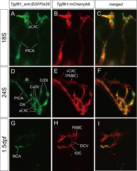
Arterial and venous characteristics of each ocular vessel. Light-sheet microscopy of the double Tg(flt1_enh:EGFP)k28 and Tg(flk1:mCherry)k6 zebrafish embryos at 18S (A-C), 24S (D-F), and 1.5 dpf (G-I). Tg(flt1_enh:EGFP)k28 (A, D, and G), Tg(flk1:mCherry)k6 (B, E, and H), and merged (C, F, and I) images. Dorsal (A-C), rostral-lateral (D-F), and lateral (G-I) views. Only the arterial components of the ocular vasculature, aCAC, OA, and NCA, expressed EGFP, whereas all vessels expressed mCherry. Asterisk in D indicate the PMBC which did not express EGFP. |

Selected images of serial sections before the reconstruction. Selected images of the serial sections at 1 (A-C) and 2 (D-G) dpf. Frontal (A-C) and horizontal (D-G) planes. Each ocular vessel and the positioning number of each selected image were indicated. |