FIGURE SUMMARY
Title

Mind Bomb-Binding Partner RanBP9 Plays a Contributory Role in Retinal Development

Authors
Yoo, K.W., Thiruvarangan, M., Jeong, Y.M., Lee, M.S., Maddirevula, S., Rhee, M., Bae, Y.K., Kim, H.G., Kim, C.H.
Source
Full text @ Mol. Cells

Spatiotemporal expression of ranbp9 mRNA during embryonic development.

Whole-mount in situ hybridization was performed with DIG-labeled antisense RNA probe (hpf, hours after fertilization). (A) At 2-cell stage, maternal transcripts were detected in high density. (B) At dome stage (4 hpf), high level of transcription persisted. (C) At the beginning of gastrulation, shield stage (6 hpf), ranbp9 mRNA was hardly detected. (D) At 6-somite stage (12 hpf), ranbp9 was seen expressed ubiquitously. (E) At 48 hpf, ranbp9 expression was mainly observed in the head region. (F) Section of head region at 72 hpf, showing ranbp9 expression in the brain (B) and eye (L, lens). Its expression is more prominent in forming layers (arrow head) of the retina, compared to the periphery (arrow). O, oral cavity.

EXPRESSION / LABELING:
Gene:
Fish:
Anatomical Terms:
Stage Range: 2-cell to Protruding-mouth

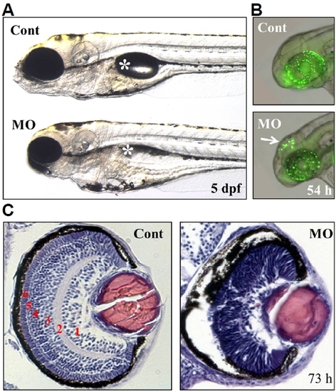
Defects in the brain and retina of ranbp9-MO injected zebrafish embryos.

(A) Control (Cont) and ranbp9-MO injected (MO) zebrafish larvae at 5 dpf. Compared to normal development of body trunk region, reduced size of brain and eye was observed in the MO injected larva. Asterisk indicates swim-bladder. (B) By acridine orange staining, a specific cell death was detected in the brain tectum of 54 hpf morphants. (C) Disruption of retinal development in ranbp9 morphant. Section of HE-stained retina in control and ranbp9 morphant at 73 hpf. In wild-type, the retina was established into several layers; 1, ganglion cell layer: 2, inner plexiform layer: 3, inner nuclear layer: 4, outer plexiform layer: 5, outer nuclear layer: and 6, photoreceptor cell layer, respectively. In morphants, however, these layer formations were not well established.

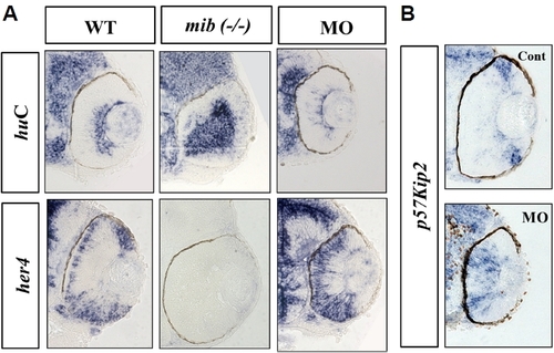
Molecular characterization of retinal development in ranbp9 morphants.

(A) Whole-mount in situ hybridization analysis for the expression of neural markers; huC for neuronal differentiation marker and her4 for neural progenitor marker, respectively. The neurogenic mutant mind bomb (mib, −/−) was used for comparison. In the WT larvae at 48 hpf, huC was expressed in the apical cells of the retina, but her4 was expressed in the basal cells. In the mib mutant larvae, huC was seen stained in most of the retinal cells near the apico-basal axis, but her4-positive cells were not observed. In the ranbp9 morphant larvae, huC expression domain was significantly reduced, however, the her4 expression domain was expanded. (B) Altered-regulation of p57kip2, a molecular marker for cell cycle exit, in ranbp9 morphants. p57kip2 transcripts were observed in the peripheral marginal regions of wild-type retina, while its expression was expanded to the central region of retina in ranbp9 morphants.

EXPRESSION / LABELING:
Genes:
Fish:
Knockdown Reagent:
Anatomical Terms:
Stage: Long-pec
PHENOTYPE:
Fish:
Knockdown Reagent:
Observed In:
Stage: Long-pec

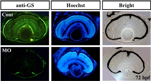
Characterization of glial cells in ranbp9 morphants.

Retinal sections were stained with an antibody against Glutamine synthetase, which is used as a molecular marker for the Müller glia cells. Glial cell differentiation is also affected in ranbp9 morphants (MO), compared to that of control retina. Hoechst staining and bright field image were shown for the comparison of retinal structures.

PHENOTYPE:
Fish:
Knockdown Reagent:
Observed In:
Stage: Protruding-mouth
Acknowledgments
This image is the copyrighted work of the attributed author or publisher, and ZFIN has permission only to display this image to its users. Additional permissions should be obtained from the applicable author or publisher of the image. Full text @ Mol. Cells