
- Title
-
Reduced DOCK4 expression leads to erythroid dysplasia in myelodysplastic syndromes
- Authors
- Sundaravel, S., Duggan, R., Bhagat, T., Ebenezer, D.L., Liu, H., Yu, Y., Bartenstein, M., Unnikrishnan, M., Karmakar, S., Liu, T.C., Torregroza, I., Quenon, T., Anastasi, J., McGraw, K.L., Pellagatti, A., Boultwood, J., Yajnik, V., Artz, A., Le Beau, M.M., Steidl, U., List, A.F., Evans, T., Verma, A., Wickrema, A.
- Source
- Full text @ Proc. Natl. Acad. Sci. USA

In vivo suppression of dock4a in a zebrafish model leads to dysplastic erythroid cells. (A) Shown are representative embryos following staining at 48 hpf with o-dianisidine to detect hemoglobin (red stain). (Top) Embryos were injected with 5 ng of standard control MO. Arrows indicate blood circulation through the caudal hematopoietic tissue, cardinal vein, and heart, with substantial staining in the sinus venosus on the yolk sac. (Lower) Embryos were injected with 5 ng of the dock4a SB MO, and at 48 hpf, they lack blood circulation and appear relatively pale with less blood in the yolk sac (n = 30 embryos for both panels). (B) Relative percentages of erythroid cells in zebrafish embryos were determined by flow cytometry comparing dock4a knockdown with control samples (with the later set to 100%). Use of gata1:dsred transgenic zebrafish embryos allowed direct assessment of erythroid cells in samples of dissociated whole embryos’ blood (n = 4 independent experiments, minimum of 200 embryos per experiment). (C) Photomicrographs of benzidine- and hematoxylin-positive erythroid cells from cytospins prepared using cells from dissociated zebrafish embryos. (Scale bars, 5 μm.) (D) BF and fluorescence images of RFP-expressing erythroid cells depicting circular- and irregular-shaped morphology harvested from embryos with reduced expression of Dock4a. (E) Quantitation of RFP-positive embryonic zebrafish blood cells that are circular or irregular in shape based on analysis of BF and RFP images using the ImageStreamX instrument. KD, knockdown. (F) Percentages of circular- and irregular-shaped erythroid cells harvested from dissociated zebrafish embryos 48 hpi of dock4a SB MO or control MO or from uninjected embryos (**P < 0.005, Student’s t test). PHENOTYPE:
|

Dock4a is expressed during zebrafish embryogenesis. Gene expression was determined using in situ hybridization. Wild-type zebrafish embryos were harvested and fixed at the stages indicated, from the 1-cell stage to 48 hpf. Embryos were hybridized to either a sense (control) probe or an antisense digoxygenin labeled RNA probe. Embryos were photographed with lateral or dorsal views as indicated (sense is a lateral view). |
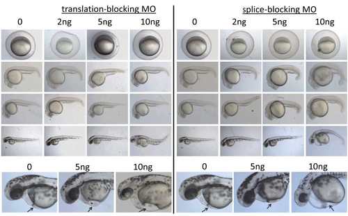
Both the TB and SB MOs cause similar embryonic defects. Wild-type zebrafish embryos were injected at the one cell stage with 0, 2, 5, or 10 ng TB (left panels) or SB (right panels) morpholino. Shown are representative embryos (n>100). Panels below are higher magnification views demonstrating a pericardial edema and the location of blood pooling in the ventral part of the embryo (arrows) PHENOTYPE:
|

Primitive hematopoiesis initiates normally in the dock4a morphant embryos. Wild-type zebrafish embryos were injected with 0 (control), 5 ng, or 10 ng of the dock4a SB morpholino. The embryos were harvested and fixed in 4% PFA at the 5, 10, or 20 somite stage. Gata1 transcript patterns were detected using an antisense digoxygenin labeled RNA probe, seen initially as two stripes in the lateral mesoderm, converging at the intermediate cell mass in the tail. EXPRESSION / LABELING:
|