
- Title
-
A role for Gle1, a regulator of DEAD-box RNA helicases, at centrosomes and basal bodies
- Authors
- Jao, L.E., Akef, A., Wente, S.R.
- Source
- Full text @ Mol. Biol. Cell
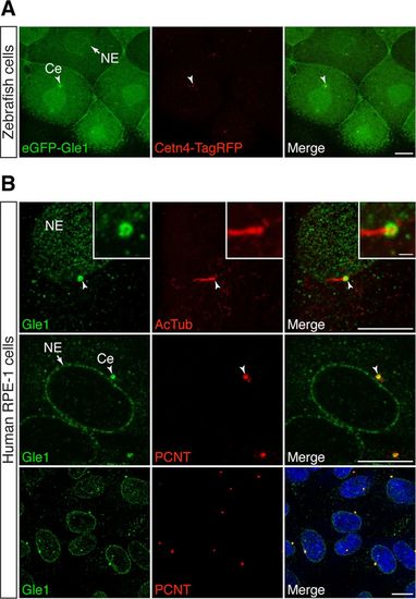
Gle1 is localized to the centrosome and basal body. (A) eGFP-tagged human Gle1 and Cetn4-TagRFP were transiently expressed from the injected RNA in the cells of dome-stage zebrafish embryos. eGFP-Gle1 localized to the NE (arrow) and the centrosome (Ce; arrowhead). (B) Confocal images of serum-starved human RPE-1 cells stained with antibodies against Gle1 and acetylated tubulin (AcTub; top) or Gle1 and PCNT (middle and bottom). Endogenous Gle1 localized to the ciliary base (top; arrowhead) and the centrosome (middle and bottom; arrowhead) as well as to NE (arrow). Scale bars: 10 μm (main images), 1 μm (insets). |
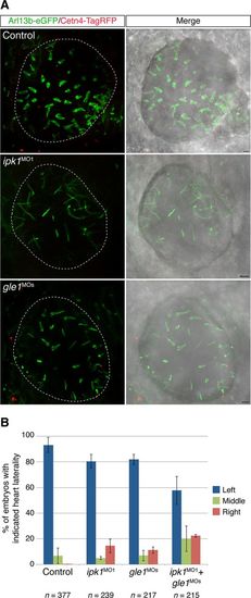
Depletion of Gle1 reduces ciliary beating and alters left–right asymmetry in zebrafish. (A) The axoneme and the basal body of the cilia were mosaically labeled by Arl13b-eGFP and Cetn4-TagRFP, respectively, in uninjected, ipk1MO1-injected, or gle1MOs-injected embryos. Motile cilia in the Kupffer’s vesicle were imaged at six- to nine-somite stages from the dorsal side, anterior to the right. Normal ciliary beating is detected by a fan-like morphology in the control. Scale bar, 5 μm. (B) Tg(-5.1mnx1:TagRFP) embryos were injected with suboptimal doses of translation-blocking ipk1 morpholino (ipk1MO1, 4 ng), a mix of two nonoverlapping translation-blocking gle1 morpholinos (gle1MOs, 1.5 ng each), or a combination of both ipk1MO1 and gle1MOs at the one-cell stage. The direction of the heart looping (TagRFP+) was assessed at 60–72 h post fertilization. Three independent injections were performed for each condition. Values are mean ± SEM; n is the number of embryos analyzed in each condition. PHENOTYPE:
|