
- Title
-
PPFIA1 drives active α5β1 integrin recycling and controls fibronectin fibrillogenesis and vascular morphogenesis
- Authors
- Mana, G., Clapero, F., Panieri, E., Panero, V., Böttcher, R.T., Tseng, H.Y., Saltarin, F., Astanina, E., Wolanska, K.I., Morgan, M.R., Humphries, M.J., Santoro, M.M., Serini, G., Valdembri, D.
- Source
- Full text @ Nat. Commun.
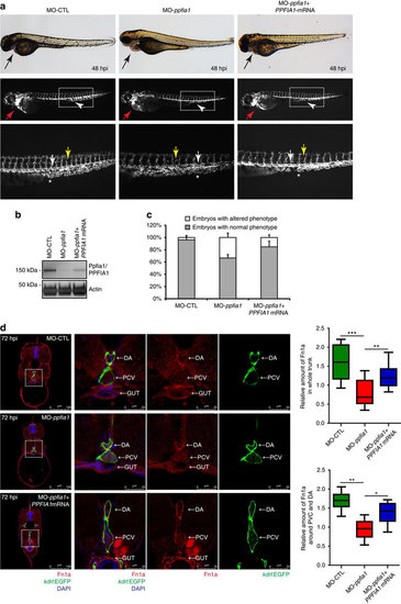
PPFIA1 silencing affects vascular morphogenesis in developing zebrafish embryo. (a) Lateral view at conventional light and confocal fluorescence microscopy of Tg(kdrl:EGFP) zebrafish embryos carrying EC-specific EGFP expression and derived from fertilized zebrafish eggs that were injected with 83 μM of a ppfia1 translation blocking morpholino (MO-ppfia1), 5-base mismatch (MO-CTL), or ppfia1 translation blocking morpholino and 40 pg PPFIA1 mRNA at the single-cell stage. Starting from 48 hpi, ∼35% of the ppfia1 morphants displayed: (i) an enlarged heart chamber phenotype (red arrows) due to atrium dilation associated with pericardial oedema, blood stasis and reduced blood flow despite the presence of cardiac activity; (ii) malformations in the intersegmental vessels (Se, yellow arrow); (iii) an irregular shape (margin) in the DA and the PCV (white arrows). Human PPFIA1 mRNA co-injection successfully rescued the cardiovascular defects in the vast majority (∼84%) of ppfia1 morphants that appeared morphologically normal (b). Western blot analysis of MO-ppfia1, MO-CTL and MO-ppfia1+PPFIA1 mRNA morphants, at 48 hpi, revealed that the band corresponding to zebrafish ppfia1 present in MO-CTL, completely disappear in MO-ppfia1 and it is partially restored in MO-ppfia1+PPFIA1 mRNA morphants. Actin was used as loading control. (c) Relative percentage of the embryos having normal or altered phenotype in two independent experiments is reported in the graph and both absolute numbers with relative percentage of the embryos for each experimental group are reported in Supplementary Tables 1 and 2. (d) Confocal fluorescence microscopy analysis of MO-CTL, MO-ppfia1 and MO-ppfia1+ PPFIA1 mRNA Tg(Kdrl:EGFP) zebrafish embryos trunk cross-sections at 72 hpi stained with DAPI and Fn1a. Right panels are magnifications of boxed areas of the left panels. While MO-CTL morphants show normal Fn1a expression around the PCV, DA, gut and in the whole trunk cross-section (upper panels); MO-ppfia1 morphants display a decreased Fn1a expression in the overall trunk region (middle panels). Co-injection of human PPFIA1 mRNA in MO-ppfia1 morphants rescued Fn1a deposition defects albeit not completely in the overall trunk cross-section. Relative amount of Fn1a expression in MO-CTL, MO-ppfia1 and MO-ppfia1+ PPFIA1 mRNA Tg(Kdrl:EGFP) zebrafish whole trunk cross-sections or around PVC and DA, at 72 hpi. Data are mean values±s.d. from 4 (MO-CTL) or 9 (MO-ppfia1 and MO-ppfia1+ PPFIA1 mRNA) embryos from two independent experiments, and 5th/95th percentile.*P<0.05; **P<0.01; ***P<0.001; ANOVA followed by the Tukey range test. Scale bar, 100 μm (d, left panels), 25 μm (d, right panels). EXPRESSION / LABELING:
PHENOTYPE:
|