
- Title
-
Identification of novel MYO18A interaction partners required for myoblast adhesion and muscle integrity
- Authors
- Cao, J.M., Cheng, X.N., Li, S.Q., Heller, S., Xu, Z.G., Shi, D.L.
- Source
- Full text @ Sci. Rep.

Analysis by in situ hybridization of myo18ab, lurap1, p190RhoGEF and Golgin45 expression pattern at 24 hpf. (A,B) Expression of myo18ab is localized at somite borders and in the somites. Diffuse expression can be observed in the head region. It is strongly localized in deep muscle and weakly in superficial muscle and in the notochord. (C,D) Expression of lurap1 is mainly localized to the head region and weakly in trunk and posterior somites. A punctate pattern can be detected in the dorsal region of the neural tube (arrows). It is also detected in the notochord. (E,F) Expression of p190RhoGEF can be detected in the head region, in trunk and posterior somites, and in the notochord. Hybridization signal can be also observed in the yolk sac. (G,H) Expression of Golgin45 is localized to the head region, and is also detected in the entire somites and notochord. nt, neural tube; nc, notochord; s, somites. Scale bars: (A,C,E,G) 350 μm; (B,D,F,H) 50 μm. EXPRESSION / LABELING:
|
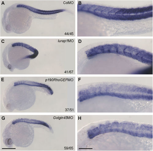
Expression pattern of muscle-specific mhc in representative control and morphant embryos at 24 hpf. (A,B) A CoMO-injected embryo showing regular mhc expression pattern in the somites. (C,D) A lurap1MO-injected embryo with bent axis and disrupted mhc expression pattern in the somites. (E,F) A p190RhoGEFMO-injected embryo with shortened anteroposterior axis and absence of mhc expression at somite boundaries. (G,H) A Golgin45MO-injected embryo with reduced anteroposterior axis associated with disrupted mhc expression pattern in the somites. Scale bars: (A,C,E,G) 350 μm; (B,D,F,H) 120 μm. PHENOTYPE:
|
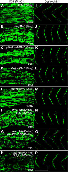
Functional interaction between MYO18A, Lurap1, p190RhoGEF and Golgin45 in myofiber integrity and dystrophin localization. Representative images showing the immunostaining results of slow muscle-specific MHC (A–H) and dystrophin (I–P) in control and various morphant embryos, as indicated. (A,I) Regular organization of myofibers within the somite (A) and localization of dystrophin at the sarcolemma (I) in CoMO-injected embryos, with clear chevron-shape myosepta. (B–E, J–M) Individual knockdown using high amounts of MOs against lurap1 (B,J), p190RhoGEF (C,K), Golgin45 (D,L) or myo18ab (E,M) produces similar muscle lesions (B–E) and disrupts dystrophin localization (J–M) at the sarcolemma. (F–H, N–P) Simultaneous knockdown using low amounts of indicated MOs also affects myofiber integrity (F–H) and dystrophin localization (N–P), with severely disorganized myofibers and strongly disrupted myosepta in a high proportion of morphant embryos. Scale bar: 100 μm. |

Knockdown of myo18ab disrupts the integrity of deeply located fast muscle cells. (A,B) Immunofluorescence staining of fast myofibers by F310 antibody in CoMO- and myo18abMO-injected embryos. (C,D) Transverse histological sections showing the organization of deeply located fast muscle cells in control and myo18abMO-injected embryos at 96 hpf. Notice that the control embryo has myofibers with centrally located nuclei, while the morphant embryo shows disorganized myofibers with nuclei located at the periphery. nt, neural tube; nc, notochord; s, somite. Scale bars: (A,B) 100 μm; (C,D) 50 μm. |

MYO18A and its interaction partners are required for adhesion of muscle cells on ECM. Myoblasts were taken from the somites of indicated control or morphant embryos at 24 hpf, and cultured on laminin substrate for 12 and 24 hours. (A-B') CoMO-injected myoblasts efficiently adhere and extensively elongate to form multinucleated cells on laminin-coated substrate. (C-D') myo18a (myo18aa and myo18ab) morphant myoblasts exhibit strongly reduced adhesion at 12 hours, and decreased ability to form elongated and multinucleated myofibers at 24 hours. (E-F') lurap1 morphant myoblasts adhere less efficiently to laminin-coated substrate at 12 hours, and elongate at a reduced extent. (G-H') p190RhoGEFMO severely impairs myoblast adhesion, morphology and formation of elongated myofibers. (I-J') Golgin45MO strongly affects myoblast adhesion at 12 hours, with few cells elongated at 24 hours. (K) Statistical analysis of elongated myoblast cells. Numbers on the top of each column indicate total cells scored from two independent experiments. (L,M) Positive MF20 immunostaining of cultured myoblast cells from CoMO- and myo18aMO-injected embryos. Scale bars: (A–J) 100 μm; (B',D',F',H',J') 50 μm; (L,M) 50 μm. PHENOTYPE:
|

MYO18A and its interaction partners are required for maintaining the morphology of the Golgi and the organization of F-actin network. Staining of the Golgi apparatus by GM130 antibody, F-actin by TRITC-phalloidin and cell nuclei by DAPI was performed in myoblasts cultured for 24 hours in laminin-coated substrate. (A-D') Localization of the juxtanuclear cis-Golgi and the organization of F-actin bundles in CoMO-injected cells. (E-H') Knockdown of myo18a (myo18aa and myo18ab) reduces the intensity of GM130 labelling in juxtanuclear cis-Golgi and leads to a disrupted F-actin organization (arrows in G,H). (I-L') Knockdown of lurap1 leads to diffuse GM130 staining and disrupts F-actin bundles within the cells, resulting in phalloidin staining accumulated at the periphery. (M-P') Diffuse GM130 staining and strongly reduced F-actin bundles in p190RhoGEF morphant myoblast cells. (Q-T') Knockdown of Golgin45 severely reduces the formation of the Golgi apparatus and F-actin bundles. Scale bars: (A–D,E–H,I–L,M–P,Q–T) 20 μm; (D',H',L',P'T') 10 μm. PHENOTYPE:
|

Knockdown of myo18ab, lurap1, p190RhoGEF and Golgin45 affects embryonic development. (A-D) Representative phenotypes from control and indicated morphant embryos at 48 hpf. (E) Statistical analysis of different phenotypes following injection of various MOs, as indicated. Numbers on the top of each column indicate total embryos blindly scored from four independent experiments. Scale bar: (A-D) 250 μm. |

Disorganization of myosepta and sarcomere structure in lurap1, p190RhoGEF and Golgin45 morphants. (A-D) Differential interference contrast images of myofiber organization in a CoMO-, a lurap1aMO-, a p190RhoGEFMO- and a Golgin45MOinjected embryo at 96 hpf, as indicated. Scale bar: 20 μm. PHENOTYPE:
|

Dose-dependent effect of different MOs on myofiber organization. (AE) Injection of a low amount (5 ng) of each of the indicated MOs mildly affects somite structure and myofiber integrity. Scale bar: 100 μm. |

Knockdown of lurap1, p190RhoGEF and Golgin45 affects the integrity of deep muscle cells. (A-D) Histological sections of indicated embryos at 96 hpf showing deep fast muscle myofiber organization. Scale bar: 50 μm. PHENOTYPE:
|

Movement defects in lurap1 and p190RhoGEF morphant embryos. Test of “escape response” on indicated embryos at 72 hpf. |