
- Title
-
Mespaa can potently induce cardiac fates in zebrafish
- Authors
- Deshwar, A.R., Onderisin, J.C., Aleksandrova, A., Yuan, X., Burrows, J.T., Scott, I.C.
- Source
- Full text @ Dev. Biol.

mesp over-expressing cells are capable of differentiating into myocardium, endocardium, vascular endothelium and facial muscle. (A) Schematic detailing transplantation experiments to assess the potential of mesp over-expressing cells. (B) Percent of host embryos with GFP-positive cells in the heart when different mesp family members were over-expressed in the transplant assay. (C-H) Host embryos at 48hpf generated from animal cap transplants of rhodamine dextran labeled myl7:EGFP donor cells of WT (C-E) or mespaa over-expressing cells (F-H). (I) Percent of host embryos with GFP-positive cells in the endocardium and vasculature or vasculature alone when different mesp family members were over-expressed in the transplant assay. (J-O) Host embryos at 48hpf generated from animal cap transplants of rhodamine dextran labeled kdrl:EGFP donor cells of WT (J-L) or mespaa over-expressing cells (M-O). E indicates endocardial cells, V designates vascular endothelial cells. (P) Percent of host embryos with GFP-positive cells in the head when different mesp family members were over-expressed. (Q-V) Host embryos at 48hpf generated from animal cap transplants of rhodamine dextran labeled acta1:EGFP donor cells of WT (Q-S) or mespaa over-expressing cells (T-V). |

Mespaa is required early in gastrulation for pro-cardiac activity but over-expressing cells do not exhibit unique cellular behaviors until after its completion. (A) Schematic outlining the strategy for exploring the temporal requirement for mespaa in pro-cardiac activity. (B) Percent of host embryos with GFP+ cells at 48 hpf after animal cap transplantation of ecr-mespaa-Δnls over-expressing cells with tebufenozide addition at different time points. Data are represented as means±SEM. (C-F) Time-lapse images of animal cap transplants of mespaa and mespab over-expressing cells together at different stages in development. |

mespaa over-expressing cells do not induce expression of a gata5-EGFP reporter until after gastrulation. (A-D) Host embryos with transplanted WT or mespaa over-expressing donor cells (gata5:EGFP, labeled with rhodamine dextran) at the bud stage (A-B) and the 7 somite stage (12 hpf) (C-D). (E-J) FACS plot of rhodamine dextran-labeled gata5:EGFP donor cells from host embryos at different stages. Cells were either WT (E,G,I) or mespaa over-expressing (F,H,J). Percentage of cells in each bin (GFP +′ve vs ′ve) is shown. |

mespaa mutant embryos do not display defects in cardiac specification but exhibit abnormalities in left-right asymmetry. (A) Schematic of the mespaahsc11 null allele that was generated. (B-C) Whole embryo morphology of WT and mespaahsc11 null mutants at 48 hpf. (D-E) nkx2.5 expression in WT and mespaahsc11 null embryos at 16 hpf. (F-G) myl7 expression in WT and mespaahsc11 null embryos at 48hpf. (H-I) Normal and reversed heart looping demonstrated with myl7 expression at 48 hpf. A demarcates the atrium and V indicates the ventricle. (J) Percent of embryos displaying normal or reversed heart looping when deficient for different Mesp family members alone or in combination when compared to WT. (K-N) The four categories of lefty1/2 expression observed in WT and Mesp deficient embryos. (O) Percent of embryos displaying the different lefty1/2 expression patterns in WT and Mesp deficient embryos. EXPRESSION / LABELING:
PHENOTYPE:
|

Pro-cardiac activity of Mespaa maps to both N- and C-terminal portions of the protein. (A) Schematic comparing the protein sequence of all four zebrafish Mesp family members. (B) Schematic of various Mesp fusion and mutant constructs and the ability of these constructs when RNA was over-expressed to confer pro-cardiac activity to transplanted cells in the animal cap. |
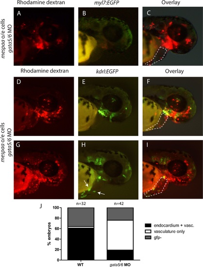
Mespaa sits upstream of gata5/6 with respect to cardiac, but not endothelial, development. (A-C) Animal cap transplants of myl7:EGFP donor embryos over-expressing mespaa and co-injected with gata5/6 MOs. (D-J) Animal cap transplants of kdrl:EGFP mespaa over-expressing cells co-injected with gata5/6 MOs. Area of the heart is outlined with a dashed line. Arrows indicate kdrl:EGFP-positive cells in the heart. Quantification of contribution is provided in (J). |

gata5 and gfp are expressed in the same pattern in gata5:EGFP transgenic embryos. (A-F) gata5 and gfp expression in gata5:EGFP embryos at 6, 10 and 14 hours post fertilization. Embryos are viewed from the animal cap, lateral and dorsal sides. (G) gata5:EGFP transgenic embryo at 6 hpf (shield stage). M indicates cells at the embryonic margin while Y indicates the yolk syncytial layer (YSL). (H-K) mespaa, gata5, gata6 and gfp expression in animal cap transplants of mespaa over-expressing Tg(gata5:EGFP) cells in host embryos at 75% epiboly (8 hpf). Embryos are visualized from an animal cap view. EXPRESSION / LABELING:
|

Depletion of Mespab, Mespba or Mespbb does not affect cardiac formation. (A-H) myl7 expression in WT and mespaahsc11 null maternal zygotic mutant embryos at 48 hpf injected with morpholinos against mespab (B,F), mespba (C,G) or mespbb (D,H) compared to uninjected controls (A,E). (I) Percent of embryos displaying normal or reversed heart looping when deficient for different Mesp family members alone or when co-injected with morpholino resistant mRNA. (J-N) The four categories of southpaw (spw) expression observed in WT and mespaahsc11 null maternal zygotic mutant embryos at the 20-22 somite stage. (N) Percent of embryos displaying the different southpaw expression patterns in WT and mespaahsc11 null maternal zygotic mutant embryos. EXPRESSION / LABELING:
PHENOTYPE:
|
Reprinted from Developmental Biology, 418(1), Deshwar, A.R., Onderisin, J.C., Aleksandrova, A., Yuan, X., Burrows, J.T., Scott, I.C., Mespaa can potently induce cardiac fates in zebrafish, 17-27, Copyright (2016) with permission from Elsevier. Full text @ Dev. Biol.