
- Title
-
The Intracellular Signaling Molecule Darpp-32 Is a Marker for Principal Neurons in the Cerebellum and Cerebellum-Like Circuits of Zebrafish
- Authors
- Robra, L., Thirumalai, V.
- Source
- Full text @ Front. Neuroanat.

ZFIN is incorporating published figure images and captions as part of an ongoing project. Figures from some publications have not yet been curated, or are not available for display because of copyright restrictions. |
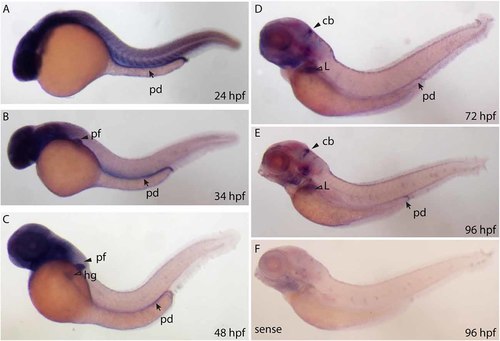
Whole mount RNA in situ hybridization using probes against the ppp1r1b-001 isoform at different stages of development. (A) 24 hours post fertilization (hpf) embryo showing ubiquitous staining including in the pronephric duct (pd). (B) More restricted staining in the central nervous sytem (CNS) and the pectoral fin buds (pf). (C) At 48 hpf, faint staining is also seen in the hatching gland (hg). (D) At 72 hpf, strong staining is seen in the cerebellum (cb), liver (L) and the pronephric duct (pd). (E) A similar staining pattern is seen at 96 hpf (F) 96 hpf larva stained with the sense probe. |
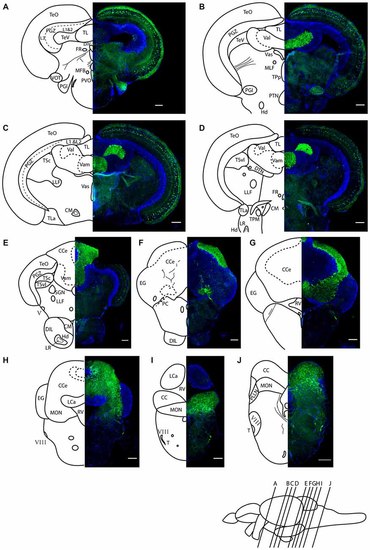
Darpp-32 immunoreactivity in adult zebrafish brain shown in rostral to caudal coronal sections. (A-J) Averaged z-stacks of serial coronal sections taken at the level and angle indicated by lines in the schematic shown in the lower right. Left hand drawings are based on the right hand image. Neuroanatomical structures are labeled according to Wullimann et al. (1996) and personal communication with M. Wullimann. DAPI: blue; anti-Darpp-32: green. Scale bar in all sections: 100 µm. Thin lines in the drawing indicate Darpp-32 immunoreactive fiber tracts. Dashed lines indicate landmarks within a neuroanatomical structure. Abbreviations used in the figure: CC, cerebellar crest; CCe, corpus cerebelli; CM, corpus mamillare; DIL, diffuse nucleus of the inferior lobe; DiV, diencephalic ventricle; DTN, dorsal tegmental nucleus; EG, Eminentia granularis; FR, habenulo-interpenduncular tract; Hd, dorsal zone of periventricular hypothalamus; LCa, caudal lobe of cerebellum; LLF, lateral longitudinal fascicle; LR, lateral recess of diencephalic ventricle; MFB, medial forebrain bundle; MLF, medial longitudinal fascicle; MON, medial octavolateralis nucleus; PC, posterior cerebellar tract; PGl, lateral preglomerular nucleus; PGZ, periventricular gray zone of optic tectum; PLLN, posterior lateral line nerve; PTN, posterior tuberal nucleus; PVO, paraventricular organ; RV, rhombencephalic ventricle; SGN; secondary gustatory nucleus; T, tangential nucleus; TeO, optic tectum; TeV, tectal ventricle; TPM, pretecto-mamillary tract; TPp, periventricular nucleus of posterior tuberculum; TL, longitudinal torus; TLa, lateral torus; TSc, central nucleus of semicircular torus; TSvl, ventrolateral nucleus of semicircular torus; Val, lateral division of valvula cerebelli; Vam, medial division of valvula cerebelli; Vas, Vascular lacuna of area postrema; ventrolateral optic tract; V, trigeminal nerve; VIII, octaval nerve. EXPRESSION / LABELING:
|
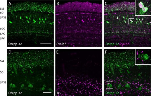
Expression of Darpp-32, Pvalb7 and tyrosine hydroxylase (TH) in the layers of the adult optic tectum. Sagittal sections of the adult optic tectum, dorsal to the top. (A-C) Colocalization of Darpp-32 and Pvalb7 immunoreactivity in somata in the SFGS and in dendrites in SM. The principal dendrite cannot always be fully seen because it does not necessary lie within the plane of the optical section shown. The layers of the optic tectum are indicated on the left hand side. Scale bar: 20 µm. However, note that the expression of Darpp-32 and Pvalb7 in the synaptic layers ventral to the SM does not overlap. (D-F) Intense TH positive fibers are visible in the vicinity of Darpp-32 expressing cell bodies. Scale bar: 50 µm. Abbreviations of the tectal layers from dorsal to ventral: SM, stratum marginale; SO, stratum opticum; SFGS, stratum fibrosum et griseum superficiale; SGC, stratum griseum central; SAC, stratum album central; SPV, stratum periventriculare. EXPRESSION / LABELING:
|
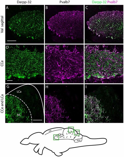
Darpp-32 and Pvalb7 expression in the cerebellum of the adult zebrafish brain. (A-C) Sagittal brain slices of the lateral division of the valvula cerebelli (Val). (D-F) Purkinje cell layer and molecular layer of the corpus cerebelli (CCe). (G-I) The caudal part of the CCe, including the lobus caudalis cerebelli (LCa). The expression of Darpp-32 and Pvalb7 overlaps in all the structures shown, albeit the intensity of expression is heterogenous. Note the restriction of expression of Darpp-32 and Pvalb7 expression to the molecular layer and Purkinje cells in (G-I). The LCa does not show any Darpp-32 staining. Scale bar in (A-C) and (G-I) is 50 µm. Scale bar in (D-F) is 20 µm. The schematic indicates the regions where the images were taken. EXPRESSION / LABELING:
|
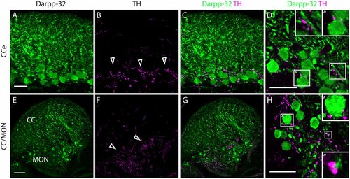
Darpp-32 and TH expression in the adult zebrafish brain. (A-C) TH-positive fibers (open arrow heads in B) run along the Purkinje cell bodies in the CCe. Collapsed z-stacks of confocal images of a coronal section. Scale bar in (A-C) 20 µm. (D) Higher magnification image showing proximity of TH puncta to Purkinje neuron somata (D′, inset size 11.09× 11.09 µm) and dendrites (D′′, inset size 9.98× 9.98 µm). Scale bar in (D) 20 µm. (E-G) Crest cells in the medial octavolateralis nucleus (MON) express Darpp-32. Their dendrites extend into the cerebellar crest (CC), Darpp-32 positive cell bodies are restricted to the MON. TH positive fibers densely innervate the MON. Mild staining in the TH channel in the CC is unspecific staining of blood vessels. Scale bar in (E-G) is 50 µm, sagittal section. (H) Higher magnification image showing TH immunoreactive puncta in close proximity to MON crest cells (H′, inset size 9.98× 9.98 µm) and dendrites (H′′, inset size 4.28× 4.28 µm), coronal section. Scale bar in (H) 20 µm. EXPRESSION / LABELING:
|
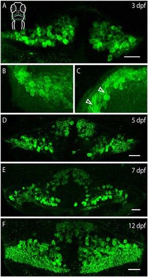
Darpp-32 expression over the course of development. Single optical sections of whole mount immunofluorescence stainings (A,D-F) and projections of a z-stack (B,C). The images are oriented as follows: (A,B,D-F) rostral up, (C) dorsal up, midline to the right. Schematic in (A) is for orientation, the images are showing the cerebellum, indicated by a green box. (A) Darpp-32 expression in the cerebellum is visible at 3 days post-fertilization (dpf). At this stage only cell bodies are visible in the dorsal view. (B) Dorsal view of a 3D projection of the left hemisphere of a 3 dpf larva. (C) Rotation of this 3D-projection around the lateral axis reveals budding dendrites growing towards the dorsal side (white arrowheads). (D-F) Darpp-32 continues to be expressed in the cell bodies and dendrites of Purkinje cells throughout the course of development. In (F) the outgrowing valvula cerebelli can be seen at rostral end of the cerebellum. Scale bar: 20 µm. EXPRESSION / LABELING:
|
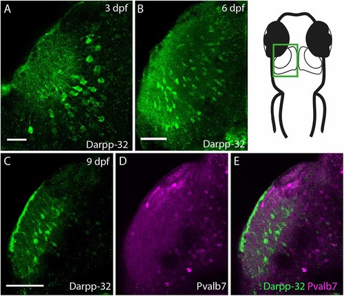
Darpp-32 expression in the larval optic tectum. Single optical sections of whole mount immunofluorescence stainings. The images are showing one hemisphere of the optic tectum as indicated in the schematic on the right side. (A) Darpp-32 expression in the optic tectum can be first seen at 3 dpf. Darpp-32 is expressed in the neuropil and the cell bodies of projection neurons. (B) Darpp-32 expression in optic tectum at 6 dpf. (C-E) Note that during the course of development the expression of Pvalb7 (D) and Darpp-32 (C) does not overlap in the optic tectum (E). In (C) intense Darpp-32 expression is visible in the superficial most layer, presumably the SM, while Pvalb 7 expression is detectable more closer to the midline, presumably in neurons of the stratum periventriculare (SPV). Darpp-32 positive cell bodies located are more lateral compared to Pvalb 7 positive ones. Scale bar in (A) 20 µm; in (B-E) 50 µm. EXPRESSION / LABELING:
|
|
Unillustrated author statements EXPRESSION / LABELING:
|