
- Title
-
Krüpple-like factors 7 and 6a mRNA expression in adult zebrafish central nervous system
- Authors
- Bhattarai, S., Sochacka-Marlowe, A., Crutchfield, G., Khan, R., Londraville, R., Liu, Q.
- Source
- Full text @ Gene Expr. Patterns

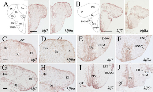
Expression of klf7 and klf6a in the anterior telencephalon and olfactory bulb of adult zebrafish brain. In the top panels, a schematic drawing of half of the brain section (dorsal side up) processed for klf7 staining is used to indicate major brain structures at this level, which is accompanied by low magnifications of half of the brain sections. Lower panels show higher magnifications of selective brain regions (same orientation) from their respective lower magnified views in the top panels. Similar arrangement is made for most of the subsequent figures. Panels C, D, G and H are higher magnifications of the dorsal halves of their corresponding images in the top panels, while panels E, F, I and J are magnified views of ventral portions of their corresponding images in the top panels. The diagonal arrow in panels A and C indicates a presumptive border region separating two brain regions with different klf7 staining. Horizontal arrowheads in panels A, C and D point to a presumptive border region in deeper brain areas with different klfs expression on either side, while vertical arrowheads in panels C and D indicate a border region in the ventricular layer with different klf staining on either side. The asterisk in panel H indicates an artificial tissue crack. See list for abbreviations. The scale bar (200 µm) in panel A applies to all images in the top panels, while the scale bar (50 µm) in panel C is for all higher magnified images. |
Expression of klf7 and klf6a in telencephalon at post anterior commissure levels. Top panels show low magnified views of halves of the brain sections, while the remaining panels show higher magnifications of the dorsal (panels C, D, G and H) or ventral (panels E, F, I and J) portions of their respective images in the top panels. See list for abbreviations. Scale bars = 200 µm for top panels, and 50 µm for the remaining panels. Images in the middle and bottom rows have the same magnifications, respectively. |
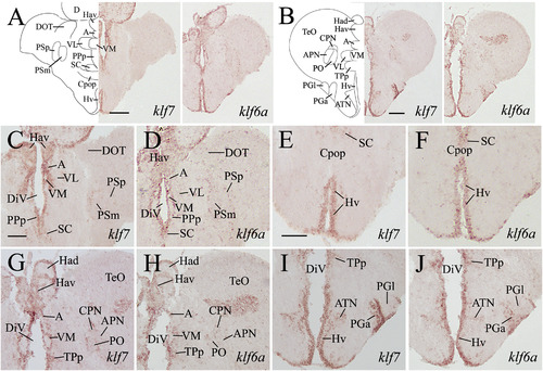
Expression of klf7 and klf6a in the preoptic, habenular, pretectal regions and diencephalon of the adult zebrafish brain. Images in the top panels are low magnified views of halves of brain sections from levels shown in Fig. 1. Panels C, D, G and H (same magnification) are higher magnifications of the dorsomedial portion of their respective images in the top panels, while panels E, F, I and J (same magnification) are ventral halves of their corresponding images in the top panels. See list for abbreviations. Scale bar = 200 µm for the top panels, 50 µm for the remaining panels. |

Expression of klf7 and klf6a in the diencephalon of the adult zebrafish brain. Top panels show low magnified views of halves of brain sections from a level shown in Fig. 1. Middle and bottom panels show higher magnifications of parts of dorsomedial and ventral diencephalon, respectively, of their corresponding images in the top panels. See list for abbreviations. Scale bar = 200 µm for the top panels, 50 µm for the remaining panels. |

Expression of klf7 and klf6a in preglomerular nuclei and hypothalamus of the adult zebrafish brain. Panels A, C, E and G show low magnifications of halves of brain sections from levels shown in Fig. 1. Panels B, D, F and H are higher magnifications of the preglomerular complex and hypothalamus of their respective lower magnified views on the left. Panels I to L show higher magnified views of more posterior hypothalamus. See list for abbreviations. All low magnified images have the same magnifications (Scale bar = 200 µm), and all higher magnified images have the same magnifications (Scale bar = 100 µm). |

Expression of klf7 and klf6a in the torus longitudinalis, optic tectum, dorsal tegmentum, isthmus, torus semicircularis and anterior medulla of the adult zebrafish brain. Images in the top panels are low magnified (same magnification, scale bar = 200 µm) views of halves of brain sections from levels shown in Fig. 1 (panel A is the same as Fig. 6E). Middle and bottom panels are higher magnified views (C and D have the same magnification, while the remaining panels have the same magnification, scale bar = 100 µm) of portions of their respective images in the top panels. Panels C and D, E and F are torus longitudinalis and optic tectum, respectively. Panels G and H, I and J show dorsomedial tegmentum and torus semicircularis, respectively. See list for abbreviations. |

Expression of klf7 and klf6a in the dorsal tegmentum, isthmus, torus semicircularis and anterior medulla of the adult zebrafish brain. Top panels show lower magnified images (scale bar = 200 µm) of halves of brain sections from a level shown in Fig. 1. Middle and bottom panels show higher magnified views (scale bar = 100 µm) of dorsomedial tegmentum and torus semicircularis, respectively. See list for abbreviations. |

Expression of klf7 and klf6a in the cerebellum and medulla of the adult zebrafish brain. Top panels show lower magnified views of halves of the brain sections from levels indicated in Fig. 1. The remaining panels are higher magnifications of the dorsomedial (C, D, G and H) or ventral portions (E, F, I and J) of the medulla of their corresponding images in the top. See list for abbreviations. Scale bar = 200 µm for the top panels, 50 µm for the remaining panels. |

Expression of klf7 and klf6a in the posterior cerebellum and middle level medulla of the adult zebrafish brain. Top panels show lower magnified images (scale bar = 200 µm) of halves of brain sections from a level shown in Fig. 1. Middle and bottom panels show higher magnified views (scale bar = 50 µm) of dorsal and ventral portions, respectively, of their corresponding images in the top panels. See list for abbreviations. |

Expression of klf7 and klf6a in the facial and vagal lobes, and the medulla of adult zebrafish. Top panels show lower magnified images (scale bar = 200 µm) of halves of brain sections from a level shown in Fig. 1. Middle and bottom panels show higher magnified views (scale bar = 50 µm) of the dorsal and ventral halves, respectively, of their corresponding images in the top panels. |
Reprinted from Gene expression patterns : GEP, 21(1), Bhattarai, S., Sochacka-Marlowe, A., Crutchfield, G., Khan, R., Londraville, R., Liu, Q., Krüpple-like factors 7 and 6a mRNA expression in adult zebrafish central nervous system, 41-53, Copyright (2016) with permission from Elsevier. Full text @ Gene Expr. Patterns