
- Title
-
EP300 Protects from Light-Induced Retinopathy in Zebrafish
- Authors
- Kawase, R., Nishimura, Y., Ashikawa, Y., Sasagawa, S., Murakami, S., Yuge, M., Okabe, S., Kawaguchi, K., Yamamoto, H., Moriyuki, K., Yamane, S., Tsuruma, K., Shimazawa, M., Hara, H., Tanaka, T.
- Source
- Full text @ Front Pharmacol

Inhibition of EP300 increases retinal apoptosis in a larval zebrafish model of light-induced retinopathy. (A) Protocol for light-induced retinal damage in larval zebrafish. Zebrafish are shielded from light between 3 and 5 days post-fertilization (dpf) and then exposed to normal conditions (14 h 250 lux/10 h dark) or intense light (13,000 lux) in the presence or absence of 2 µM C646 for 24 h at 27°C. After light exposure, whole-mount TUNEL staining was performed. (B) Representative images of TUNEL staining in the retina of zebrafish exposed to intense light (indicated as light+) or normal light conditions (light-). Scale bars, 100 µm. (C) Quantitative analysis of retinal apoptosis in zebrafish exposed to the conditions shown in (B). *p < 0.01, **p < 0.001, ***p < 0.0001. Data are the mean ± SEM of 13-14 zebrafish/group. PHENOTYPE:
|

Inhibition of EP300 reduces the photoreceptor cell outer segments in a zebrafish model of light-induced retinopathy. (A) Protocol for light-induced retinal damage in larval zebrafish, as described for Figure 4A. After light exposure, whole-mount immunohistochemical staining with anti-Zpr3 antibody was performed. (B) Representative images of anti-Zpr3 antibody staining of zebrafish exposed to normal or intense light. Scale bars, 100 µm. (C) Quantitative analysis of photoreceptor cell outer segments of zebrafish exposed to the conditions shown in (B). *p < 0.05. Data are the mean ± SEM of n = 24 zebrafish/group for light-/C646- and light-/C646+, n = 17 for light+/C646-, and n = 22 for light+/C646+. |
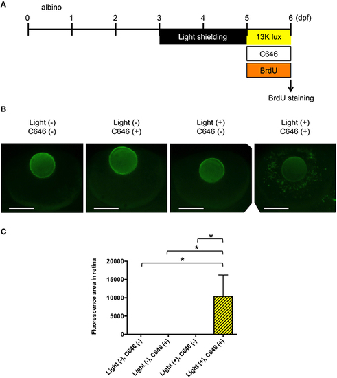
Inhibition of EP300 increases BrdU incorporation in putative Müller cells in the zebrafish model of light-induced retinopathy. (A) Protocol for light-induced retinal damage in larval zebrafish, as described for Figure 4A. After light exposure, whole-mount immunohistochemical staining with anti-BrdU antibody was performed. (B) Representative images of anti-BrdU antibody staining of zebrafish exposed to normal or intense light. Scale bars, 100 µm. (C) Quantitative analysis of BrdU-positive cells in the retinas of zebrafish exposed to the conditions shown in (B). *p < 0.05. Data are the mean ± SEM of n = 8 for all groups. PHENOTYPE:
|