
- Title
-
Extracellular interactions and ligand degradation shape the nodal morphogen gradient
- Authors
- Wang, Y., Wang, X., Wohland, T., Sampath, K.
- Source
- Full text @ Elife
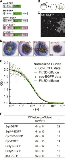
Activity range and diffusion of Nodal-GFP fusion proteins. (A) Constructs used for profiling fluorescent Nodal fusion proteins in embryos. S, signal peptide; Pro, pro-domain; Mat, mature-domain; sec-EGFP, secreted EGFP. Red arrow indicates convertase cleavage sites. (B) Injection procedure. (C) Confocal image of an injected embryo at 30% epiboly stage. White crosses mark the extracellular spots where the FCS measurements were taken. (D) Representative images of RNA in situ hybridization showing the activity range of Sqt, Cyc and mutant Nodals. Source cells are marked in brown and blue staining indicates expression of the Nodal target ntl. Scale bars, 50 µm. (E) Representative auto-correlation functions (dots) and fittings (line) of Sqt-EGFP (green) and sec-EGFP (black). (F) Table showing diffusion coefficients of the Nodal and Lefty fusion proteins as measured by FCS. |

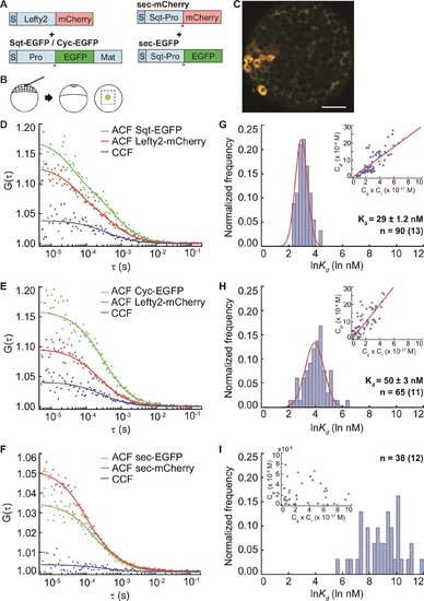
Sqt has higher affinity to the Acvr2b receptors than Cyc. (A) Sqt-/Cyc-/sec-EGFP and Acvr2b-mCherry constructs. S, signal peptide; Pro, pro-domain; Mat, mature-domain; ECM, extracellular and transmembrane domain. Red arrows indicate convertase cleavage sites. Sqt signal peptides and pro-domain were used in sec-EGFP constructs. (B) Injection procedure. (C) Representative image of an injected embryo at 30% epiboly stage showing the expression patterns of the fusion proteins. Scale bar represents 50 µm. (D,E,F) Representative auto-correlation (ACF) and cross-correlation functions (CCF) and fits. (G,H,I) Individual Ln(Kd) frequency histogram and Gaussian fitting (red curve). Inset, concentration plot and linear regression (red line). X axis, concentration of bound protein (Cgr (x10-9 M)); Y axis, products of concentrations of free proteins (Cg x Cr (x10-16 M)). n = number of data points (number of embryos). |
FCCS measurements reveal that Lefty has higher affinity to Sqt compared to Cyc. (A) Constructs used for injection. S, signal peptide; Pro, pro-domain; Mat, mature-domain. Red arrows indicate the convertase cleavage sites. (B) Injection procedure. (C) Confocal image of an injected embryo at 30% epiboly showing the expression patterns of the fusion proteins. Scale bar represents 50 µm. (D, E, F) Representative auto- and cross-correlation functions (ACF; CCF) and fittings. (G, H, I) Individual Ln(Kd) frequency histogram and Gaussian fits (red curve). Inset, concentration plot and linear regression (red line). X axis, concentration of bound protein (Cgr(x10-9 M)); Y axis, products of concentrations of free proteins (Cg x Cr(x10-17 M)). n = number of data points (i.e., number of embryos). |
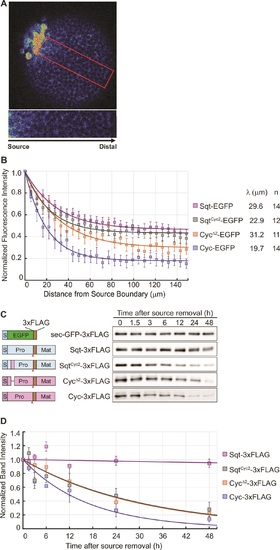
The distribution of Nodal proteins correlates with clearance. (A) Upper, representative image and region of interest (red rectangle) for measuring distribution; lower, inset showing magnified region of interest. (B) Normalized distribution profiles and fitting. Error bars indicate standard error of mean (s.e.m). (C) Representative western blots of Nodal proteins harvested from HEK293T cell culture medium at different time points after removal of the source. The Nodal proteins were immuno-precipitated with anti-FLAG antibody and detected by western blot with the same antibody. Schematics on the left show the position of the FLAG epitope tags in each construct. (D) The profile of Nodal protein levels over time after source removal. The data points were fitted with an exponential decay model. Error bars indicate s.e.m. |
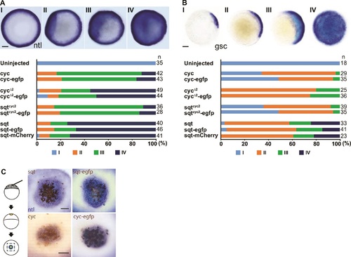
Tagged Nodals show similar activity compared to their untagged counterparts. (A) Induction of ntl in embryos overexpressing Nodal or Nodal fusions. Five picogram aliquots of RNA was injected into one-cell stage wild-type embryos and ntl transcript expression was examined at 50% epiboly. Animal pole views of embryos showing endogenous ntl expression (I), and mild (II) or massive (III and IV) expansion of the ntl expression domain. Embryos were assessed and counted accordingly. Percentages for each class are shown in the histogram. (C) Induction of gsc in embryos overexpressing Nodal or Nodal fusions. Animal pole views of embryos showing endogenous gsc expression (I), mild expansion (II) or massive expansion (III and IV) of gsc expression domains. (C) Five picogram aliquots of RNA encoding Nodal or Nodal fusions were injected into one-cell at the 128-cell stage with a lineage tracer (Biotin-Dextran, brown color staining). Range of signaling was examined by detecting ntl transcription (blue/purple color staining). Scale bars represent 100 µm. |