
- Title
-
Deguelin-induced blockade of PI3K/protein kinase B/MAP kinase signaling in zebrafish and breast cancer cell lines is mediated by down-regulation of fibroblast growth factor receptor 4 activity
- Authors
- Wu, W., Hai, Y., Chen, L., Liu, R.J., Han, Y.X., Li, W.H., Li, S., Lin, S., Wu, X.R.
- Source
- Full text @ Pharmacol Res Perspect

Growth repression and apoptosis induction caused by deguelin. (A) Morphological change in zebrafish with or without deguelin treatment. Significant growth retardation can be found in 200 and 500 nmol/L deguelin-treated group. (B) Whole-mount embryos labeled with anti-pH3 antibody to examine proliferating cells in zebrafish larvae. The numbers of pH3-positive cells decreased dramatically and rarely expressed with 200 nmol/L deguelin treatment (magnification 50×). (C) Phenotypic assessed by terminal deoxynucleotidyl transferase dUTP nick end labeling (TUNEL) staining. There was a dose-dependent increase of apoptotic cells in TUNEL assay. (magnification 50×). |
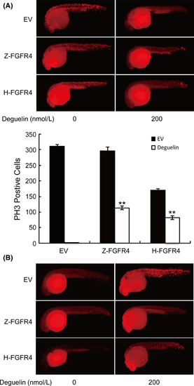
The counteractant effect of overexpressing FGFR4 in zebrafish after deguelin treatment. (A) Zebrafish embryos were labeled with anti-pH3 antibody after injection of pEGFP-C3 containing FGFR4 (Z-FGFR4 and H-FGFR4 stand for zebrafish and human FGFR4, respectively) to detect proliferating cells. DyLight 594 secondary antibody was used to avoid green fluorescence emitted by pEGFP-C3 vector. Up-regulation of FGFR4 has partly restored proliferating cells compared with the control group. PH3-positive cells are counted in Image J. **P < 0.01 (t-test) (B). TUNEL assay was conducted to analyze the apoptosis after the injection of FGFR4. Apoptotic cells were reduced in the trunk areas in injected groups. FGFR4, fibroblast growth factor receptor 4; TUNEL, terminal deoxynucleotidyl transferase dUTP nick end labeling. |