
- Title
-
GADD45B mediates podocyte injury in zebrafish by activating the ROS-GADD45B-p38 pathway
- Authors
- Chen, Z., Wan, X., Hou, Q., Shi, S., Wang, L., Chen, P., Zhu, X., Zeng, C., Qin, W., Zhou, W., Liu, Z.
- Source
- Full text @ Cell Death Dis.
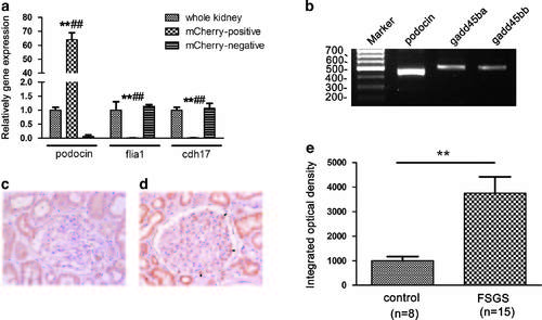
Validation of gadd45ba/bb expression on podocyte. (a) qRT-PCR validation of isolated podocytes. qRT-PCR analysis confirmed that the expression of podocyte marker podocin in isolated podocytes (mCherry positive) was highly enriched as compared with those from whole kidney and mcherry-negative cells. In contrast, expression of endothelial marker flia1 and tubular marker cdh17 were significantly lower in isolated podocytes as compared with those in whole kidney and mCherry-negative cells. Results are represented as mean±S.D. (n=3), **P<0.01 compared with whole kidney, ##P<0.01 compared with mcherry-negative cells. (b) RT-PCR analysis of gadd45ba/bb mRNA expression in isolated podocytes. gadd45ba/bb expressed in isolated podocytes which express podocin (c, d) GADD45B expression in biopsy of normal control (c) and FSGS patient (d) by immunohistochemical staining. GADD45B signals are very weak in glomeruli of normal kidney tissues, whereas GADD45B expression in FSGS patients was significantly increased as compared with normal control group. Arrows indicate GADD45B expression on podocytes of FSGS patient. Original magnification × 400 (e) Semiquantitative analysis of immunohistochemical staining results showed GADD45B expression of glomeruli of FSGS patients were significantly higher than the control group. Results are represented as mean±S.D. **P<0.01 EXPRESSION / LABELING:
|

gadd45ba and gadd45bb are induced by podocyte Injury in zebrafish. (a) Dissected renal glomeruli from Tg(pod:gal4;UAS:NTR-mcherry), mCherry expression in podocytes allow for easy collection under fluorescence microscope. (b–d) Quantitation of nephrin (b), gadd45ba (c) and gadd45bb (d) transcripts by quantitative RT-PCR before and after MTZ (120 µM) treatment. Results are represented as mean±S.D. (n=3), **P<0.01 |

gadd45ba/b overexpression in podocytes aggravates MTZ-induced podocyte injury. (a) Schematic graph showing the generation of transgenic fish. Podocyte-specific expression of gadd45ba/b is confirmed by in situ hybridization in WT (A1), Tg (pod:Gal4,UAS:gadd45ba) (A2) and Tg (pod:Gal4,UAS:gadd45bb) (A3) at 3dpf. (b) Experimental scheme to test gadd45ba/b overexpression on MTZ-induced podocyte injury. (c) Representative figures showing the phenotypes due to MTZ-induced podocyte injury. (c1) Dorsal view of five dpf larvae showing periorbital edema (red arrowhead). (c2) side view of five dpf larvae showing whole-body edema (red arrowhead). (c3) Cross-section of eyes of five dpf larvae stained with methylene blue showing the severity of periorbital edema (asterisks). (c4) TEM showing the degree of foot-process effacement. (d) Quantitation of the percentage of larvae with periorbital edema. The nonfluorescence fish were used as negative controls and had no observable periobital edema phenotypes after being treated with MTZ. (e) Quantitation of the percentage of larvae with mild and severe edema. Results are represented as mean±S.D. (n=3), *P<0.05, **P<0.01 versus NTR group, #P<0.05 versus NTR and gadd45bb group EXPRESSION / LABELING:
PHENOTYPE:
|

gadd45ba and gadd45bb overexpression in podocytes aggravates MTZ-induced proteinuria. (a) Representative fluorescence figures showing disruption of the glomerular filtration barrier by MTZ leads to accumulation of GFP fluorescence in the proximal tubules (arrowheads) in Tg(pod:Gal4;UAS-NTR-mCherry; lfabp:VDBP-GFP). mCherry fluorescence (arrows) is reduced in glomeruli and accumulated in proximal tubules presumably due to podocytes loss. (b) Quantitation of GFP by ELISA showing zebrafish expressing gadd45ba/b in podocytes exhibit more poteinuria induced following MTZ-induced podocyte injury. Results are represented as mean±S.D. (n=3), *P<0.01 versus NTR group; Δ P<0.01 versus NTR and gadd45bb group |

gadd45ba/gadd45bb overexpression aggravated podocyte apoptosis. Confocal images of the pronephric glomeruli in zebrafish embryos were treated with 100 µM MTZ from 72 hpf to 96 hpf. Green fluorescence represents active caspase-3 staining; podocytes are marked with mCherry red fluorescence and nuclei are labeled with DAPI (original magnification, × 630) |

ZFIN is incorporating published figure images and captions as part of an ongoing project. Figures from some publications have not yet been curated, or are not available for display because of copyright restrictions. PHENOTYPE:
|
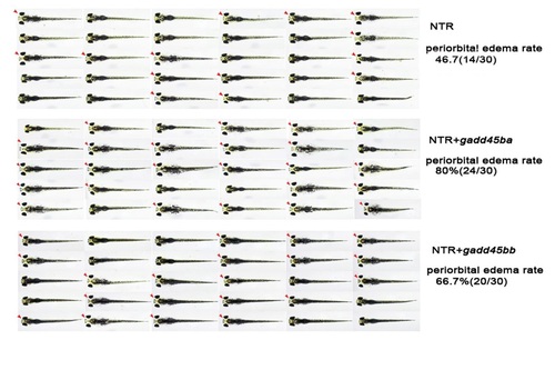
Figures of each individual 5 day-old larva following 100 µM MTZ treatment for 48 hours showing the occurrence of periorbital edema ( Arrowhead mark larvae with periorbital edema). |