
- Title
-
The Effects of Hsp90α1 Mutations on Myosin Thick Filament Organization
- Authors
- He, Q., Liu, K., Tian, Z., Du, S.J.
- Source
- Full text @ PLoS One
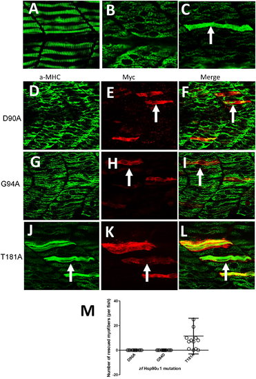
Mutating D90, G94 but not T181 in the N-terminal ATP binding domain disrupts Hsp90α1 function in myosin thick filament organization. DNA construct expressing the myc-tagged wild type Hsp90α1, D90A, G94D or T181A mutant was co-injected with Hsp90α1 ATG-MO into fertilized eggs of zebrafish. The injected embryos were analyzed by double staining with anti-myc (9E10) and anti-MHC (F59) antibodies at 28 hpf. A-C. Anti-MHC antibody staining shows the thick filament organization in skeletal muscles of control (A), Hsp90α1 knockdown (B), or DNA and ATG-MO co-injected (C) embryos. D-F. Myosin thick filament organization and myc-tagged D90A expression in skeletal slow muscles of a zebrafish embryo co-injected with Hsp90α1 ATG-MO and D90A construct. G-I. Myosin thick filament organization and myc-tagged G94D expression in skeletal slow muscles of a zebrafish embryo co-injected with Hsp90α1 ATG-MO and G94D construct. J-L. Myosin thick filament organization and myc-tagged T181A expression in skeletal slow muscles of a zebrafish embryo co-injected with Hsp90α1 ATG-MO and T181A construct. M. Plot showing the number of rescued myofibers in 12–13 individual zebrafish embryos injected with D90A, G94D or T184A mutant. Scale bar = 20 μm. |

Thr33 phosphomimetic mutation blocks Hsp90α1 function in thick filament organization. DNA construct expressing the myc-tagged T33A/Y35F double, or T33E and T33D single mutant was co-injected with Hsp90α1 ATG-MO into fertilized eggs of zebrafish. The injected embryos were stained with anti-myc (9E10) and anti-MHC (F59) antibodies at 28 hpf. A-C. A mosaic and cell-autonomous pattern of rescue was detected in myofibers expressing the non-phosporylatable T33A/Y35F mutant. D-I. Expression of the phosphomimetic T33D (D-F) or T33E (G-I) mutant failed to rescue the myosin thick filament defect in skeletal muscles. H. Plot showing the number of rescued myofibers in 10–16 individual embryos injected with T33A/Y35F, T33E or T33D mutant. A few fibers showed a partial rescue for T33E. Scale bar = 20 μm. |

The varied effect of T87 mutation on Hsp90α1 function in myofibril organization. DNA construct expressing the non-phosporylatable T87A mutant or phospho-mimic T87E mutant was co-injected with Hsp90α1 ATG-MO into fertilized eggs of zebrafish. The injected embryos were double stained with anti-myc (9E10) and anti-MHC (F59) antibodies at 28 hpf. A-C. A mosaic and cell autonomous pattern of rescue was detected in all myofibers expressing the non-phosporylatable T87A mutant. D-I. Expression of the phospho-mimic T87E mutant resulted in a varied rescue on myosin thick filament organization. 14% (n = 35) of the T87E expressing myofibers failed to show any sign of rescue in myosin thick filament organization (D-F). In contrast, 39.5% (n = 100) and 46.5% (n = 117) of the T87E expressing slow myofibers showed a partial (not shown) or a full (G-I) rescue, respectively. |

K287Q acetylation mimicking mutation affects Hsp90α1 function in thick filament organization. DNA constructs expressing the myc-tagged K287R or K287Q mutant were co-injected with Hsp90α1 ATP-MO into fertilized eggs of zebrafish. The injected embryos were stained with anti-myc (9E10) and anti-MHC (F59) antibodies at 28 hpf. A-C. A mosaic pattern of rescue was detected in myofibers expressing the Hsp90α1 K287R mutαnt, mimicking the unacetylated lysine. D-F. K287Q mutant, mimicking the acetylated lysine state, failed to rescue the myofibril defect (D-F). G. Plot showing the number of rescued myofibers in 8–10 individual embryos injected with K287R, or K287Q mutant. Scale bar = 20 μm. |

Hypomethylation mimicking mutations at K206 and K608 have no effect on Hsp90α1 function in myosin thick filament organization. DNA construct expressing K206R or K608R mutant, mimicking the hypomethylated state of lysine residue, was co-injected with Hsp90α1 ATP-MO into fertilized eggs of zebrafish. The injected embryos were double stained with anti-myc (9E10) and anti-MHC (F59) antibodies at 28 hpf. A-F. A mosaic pattern of rescue was detected in all myofibers expressing the Hsp90α1 K206R (A-C) or K608R (D-F) mutant. * indicates a multinucleated fast myofiber expressing K608R mutant. The anti-MHC (F59) antibody is a slow fiber specific antibody which does not label MHC expressed in fast myofibers of zebrafish embryos. G. Plot showing the number of rescued myofibers in 9 individual embryos injected with K206R, or K608R mutant. Scale bar = 20 μm. |