
- Title
-
Still Heart Encodes a Structural HMT, SMYD1b, with Chaperone-Like Function during Fast Muscle Sarcomere Assembly
- Authors
- Prill, K., Windsor Reid, P., Wohlgemuth, S.L., Pilgrim, D.B.
- Source
- Full text @ PLoS One

Still heart, a smyd1b mutant, has defects in heart and fast skeletal muscle tissue. A lateral view of 48hpf wild type (A) and still heart (sth) mutants (B), which have pericardial edema, small eyes, malformed head and reduced motility. Black arrowheads highlight the pericardial edema in sth mutants and the absence of edema in wild type. White arrowheads indicate blood pooling in the mutant and the absence of pooling in wild type. (C&D) Sth mutant hearts are underdeveloped and do not beat. (E-G) Examination of lateral myofibers at 5dpf under DIC microscopy revealed striations, indicative of fully formed sarcomeres, are visible in the myofibers of wild type muscle (E, black arrowheads), while absent in the fast muscle of still heart mutants (F); striations are present in sth slow muscle (G, black arrowhead) but are disturbed by nuclei and fluid-filled spaces (G, white arrowhead). Sequencing of smyd1b cDNA from wild type embryos (H) and sth mutant embryos (I) revealed a 9 nucleotide insertion between exon 1 and 2 in the smyd1b mRNA, creating an in-frame stop codon (I, underlined sequence). The insertion is the first 9 nucleotides of intron one as sequenced from wild type smyd1b genomic sequence (J). This is a result of a transition mutation in the splice donor site of intron 1 (I, green letter in sequence, J, outlined letter in sequence). This results in a premature truncation of the SMYD1b protein after exon 1 (K). |

Myosin expression is unaffected by the absence of SMYD1b. At 24hpf, fast myosin (myhc4) expression in wild type (A) and still heart embryos (B) is similar within the trunk muscle. Slow myosin (smyhc1) expression in wild type (C) and still heart embryos (D) is also similar at 24hpf. Quantitative PCR on wild type and sth mutants for fast and slow myosin expression shows myosin expression is normal when compared to wild type embryos at 24hpf (E). (qPCR: n = 3, 30 embryos each time/phenotype. Error bars are standard deviation.). EXPRESSION / LABELING:
|
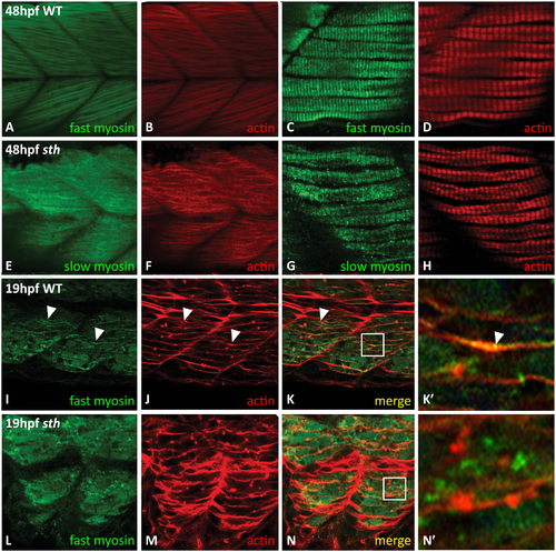
SMYD1b is required for fast myosin incorporation during sarcomere assembly. At 48hpf, fast myosin (F310—green) and actin (phalloidin—red) staining is visible in the premyofibrils in wt zebrafish tails (A&B), while absent from the premyofibrils in sth fast muscle tissue (C&D). Slow muscle (F59) develops normally in both wild type and still heart zebrafish at 48hpf (E&F, G&H). At 19hpf, in wild type embryos, fast myosin (F310) is beginning to be incorporated into the maturing myofibril (I) and overlaps (white arrowheads) with the developing actin (phalloidin) (J) fibers in trunk muscle (K, merge, K’ inset, white arrowhead). Fast myosin is not incorporated into the maturing premyofibril (L), although actin fibers are still present (M&N, N’ inset). EXPRESSION / LABELING:
PHENOTYPE:
|
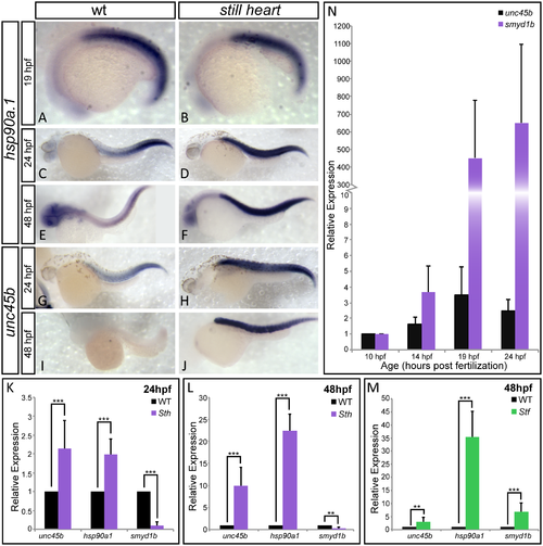
SMYD1b is co-regulated with other myosin chaperones HSP90a1 and UNC45b. In-situ hybridization staining of wild type and still heart embryos shows that hsp90a1 expression is normal in still heart mutants, when compared to wild type embryos at 19hpf (A&B). However, the expression of hsp90a1 and unc45b dramatically increases in still heart mutants at 24hpf (C-F & K) and 48hpf throughout the somites (G-J & M). Additionally, smyd1b expression increases significantly when UNC45b is absent in steif mutants (N), supporting co-regulation of these three genes. (O) A time course of smyd1b expression during muscle formation reveals that smyd1b is expressed early at 10hpf when unc45b is expressed and increases in its expression as muscle development progresses. Due to the rapid development of unc45b staining in the somites of the embryo in panel J, the head while present, has no background staining and is not clear in this focal plane. (qPCR: n = 3, 30 embryos each time/phenotype. Error bars are standard deviation.). |

Vasculature is normal in still heart mutants at 48hpf. Lateral view of 48hpf zebrafish vasculature demonstrates a repeating network of arteries and veins that highlight each somite in the trunk. Comparing vasculature organization between wild type (A) and still heart (B), there are no visible differences. (n = 10 embryos) |

Unillustrated author statements |