
- Title
-
Identification of the zebrafish red nucleus using Wheat Germ Agglutinin transneuronal tracing
- Authors
- Matsui, H., Namikawa, K., Köster, R.W.
- Source
- Full text @ Commun. Integr. Biol.

Identification of the cerebello-ruber tract in zebrafish WGA immunostaining of zebrafish brains with mosaic expression of WGA only in the right hemisphere of the cerebellum. Transient transgenic Tg(tagRFP-T:PC:WGA) zebrafish with red fluorescence only in the right cerebellar hemisphere were raised to adulthood and processed for WGA expression by immunohistochemistry (A and B). Bilateral WGA expression could be observed in the thalamus (C). Instead, expression in the descending octaval nucleus was ipsilateral to the WGA-expressing hemisphere (D), while expression in the lateral reticular nucleus (E) and red nucleus (F) were only found on the contralateral side of the neuraxis. Solid arrows: WGA signals. Dashed arrows: absence of WGA signals. n = 3. |
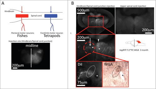
Identification of the rubro-spinal tract in zebrafish DiI tracing from the hindbrain-spinal cord junction. (A) The schematic drawing in the upper left corner illustrates the origin of motor-neurons innervating pectoral fin muscles in fish (red) and forelimb muscles in tetrapods (blue).13 (B) Red and blue arrows indicate the injection site of DiI respectively. Application of DiI into the hindbrain-spinal cord junction of transgenic Tg(tagRFP-T:PC:WGA) zebrafish labeled a contralateral neuronal nucleus containing WGA demonstrating its identity as an efferent structure of Purkinje cells (white arrow). The arrowhead points to the adjacent nucleus of the medial longitudinal fasciculus, asterisks: habenulo-interpeduncular tract. In contrast application of DiI into the upper spinal cord did not label the same WGA-positive structure. n = 5 for each injection site. |