
- Title
-
CLARITY and PACT-based imaging of adult zebrafish and mouse for whole-animal analysis of infections
- Authors
- Cronan, M.R., Rosenberg, A.F., Oehlers, S.H., Saelens, J.W., Sisk, D.M., Smith, K.L., Lee, S., Tobin, D.M.
- Source
- Full text @ Dis. Model. Mech.
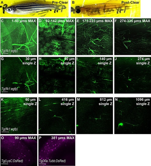
CLARITY protocol adapted for imaging intact zebrafish adults. (A,B) Zebrafish adult pre- and post-clearing. (C-N) Whole-body CLARITY allows imaging of fluorescent vasculature deep within the adult body. Blood vessels labeled by Tg(flk1:egfp) are imaged from the surface to deep within while maintaining fluorescence intensity and resolution. (C-J) Individual images obtained using an SP8 confocal microscope, ranging from the animal′s scales (surface=1µm) to 335µm deep. (C-F) Z-stack is split into ~50µm maximum projection images to allow for clear views of vascular structures. (G-J) Individual Z planes from stack. (K-N) Individual images from two-photon microscopy ranging from the animal′s scales (surface=1µm) to >1mm deep. 1-µm optical sections are shown. Scale bars: 100µm. Single Z frames were exported and gamma adjusted in FIJI/ImageJ for increased visibility, with all gamma adjustments applied uniformly across all images. (O,P) CLARITY techniques are compatible with red fluorescent proteins. (O) Neutrophils within the epidermis were imaged using the transgenic line Tg(LysC:DsRed). 90-µm maximum projection image. (P) Neuronal cell bodies within the eye of cleared zebrafish in a 381-µm maximum projection from the transgenic line Tg(Xla.Tubb:DsRed). |
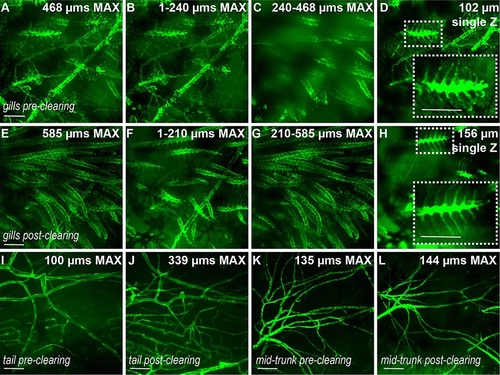
PACT protocol maintains integrity of blood vessels. (A-D) Blood vessels in gills labeled by Tg(flk1:egfp) in a zebrafish adult pre-clearing (immediately post-euthanasia) and the same animal (E-H) post-clearing. Increasing numbers of gill blood vessels are visible deeper within the body following clearing (G compared to C). Single Z frames (D,H, and insets) demonstrate that fine structures are unaffected by the clearing process. (I,J) Blood vessels in the tail of the same animal pre- and post-clearing. (K,L) Large blood vessels in the mid-trunk of the same animal pre- and post-clearing. Scale bars: 120µm. Single Z frames were exported and gamma adjusted in FIJI/ImageJ for increased visibility, with all gamma adjustments applied uniformly across all images. |
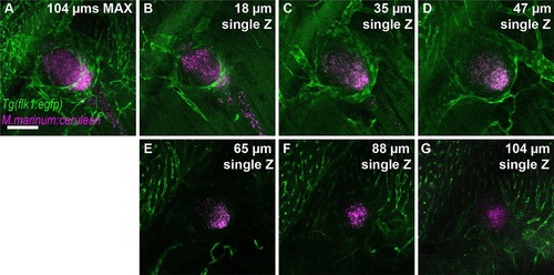
Granuloma-induced angiogenesis and mycobacterial granulomas within intact adult zebrafish. (A-G) Tg(flk1:egfp) labels blood vessels in green; magenta labels cerulean-tagged Mycobacterium marinum (Mm-cerulean), which lies within a granuloma. Imaging commences at ~400µm below the scales; stack is ~104µm deep. Scale bar: 100µm. Single Z frames were exported and gamma adjusted in FIJI/ImageJ for increased visibility, with all gamma adjustments applied uniformly across all images. |
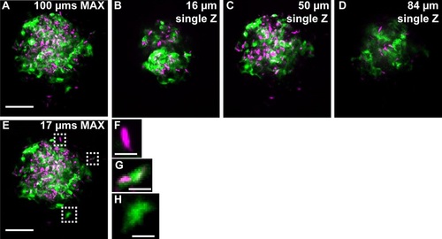
Fluorescent mycobacteria and cytokine induction can be imaged deep within intact adult zebrafish. (A) The TNF reporter (green) is expressed throughout a large granuloma (tdTomato-expressing M. marinum: magenta) in the TgBAC(tnf:GFP) line. Imaging begins 256µm below the scales and the stack (A) is µ100µm deep; individual Z planes from the stack (B-D) reveal TNF reporter intensity throughout the granuloma. (E-H) TNF reporter expression in the granuloma is not dependent on infection status of individual cells: (F) an infected cell that does not express the TNF reporter; (G) an infected cell expressing the TNF reporter; (H) an uninfected cell expressing the TNF reporter. Scale bars: 50µm (A-E); 5µm (F-H). Single Z frames were exported and gamma adjusted in FIJI/ImageJ for increased visibility, with all gamma adjustments applied uniformly across all images. |

Whole animal clearing clears internal organs. (A,B,C) Intestine dissected from PACT-cleared whole Tg(Flk1:eGFP) animal is clear (A) and blood vessels can be imaged with an epiflourescent non-confocal microscope (B,C). (B) shows a 680 µm max projection, while (C) shows a single plane at a depth of 511 µm. (D) Brain dissected from PACT-cleared whole Tg(Flk1:eGFP) animal. (E,F,G) Single planes from indicated regions of the brain boxed in (D). (H,I,J) Depth-coded maximum projection images of brain vasculature. Scale bars are 100 µm. Single Z frames were exported and gamma adjusted in FIJI/ImageJ for increased visibility, with all gamma adjustments applied uniformly across images from either top or bottom stack. |