
- Title
-
Retinoic Acid Signaling Regulates Differential Expression of the Tandemly-Duplicated Long Wavelength-Sensitive Cone Opsin Genes in Zebrafish
- Authors
- Mitchell, D.M., Stevens, C.B., Frey, R.A., Hunter, S.S., Ashino, R., Kawamura, S., Stenkamp, D.L.
- Source
- Full text @ PLoS Genet.
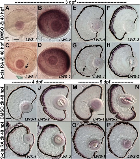
Changes in spatiotemporal patterns of expression of LWS1 and LWS2 in response to 9-cis RA treatment. A-H. Whole-mounted (A-D) and sectioned (E-H) embryo eyes obtained from embryos treated with DMSO (A,B,E,F) or 0.3 µM 9-cis RA (C,D,G,H) from 48 hpf to 75 hpf, and then hybridized with LWS1 (A,E,C,G) or LWS2 (B,G,D,H) cRNA. Arrows in C and G indicate LWS1-expressing cones in ventral retina; n, nasal; v, ventral. I-L. Sectioned embryo eyes obtained from embryos treated with DMSO (I,J) or 0.3 µM 9-cis RA (K,L) from 48 hpf to 4 dpf, and then hybridized with LWS1 (I,K) or LWS2 (J,L) cRNA. M-P. Sectioned embryo eyes obtained from embryos treated with DMSO (M,N) or 0.3 µM 9-cis RA (O,P) from 48 hpf to 4 dpf, and then hybridized with LWS1 (M,O) or LWS2 (N,P) cRNA. Scale bar in A (applies to A-H) = 50 um. Scale bar in I (applies to I-L) = 50 µm. Scale bar in M (applies to M-P) = 100 µm. |

A GFP reporter for LWS1 indicates a switch from LWS2 to LWS1 in response to RA treatment. A-C. Whole mount confocal images of retinas from LWS:PAC(H) embryos treated with DMSO (A) or 0.3 µM 9-cis RA (B) from 48 to 96 hpf. LWS2:RFP+ cones (red) are found in control retinas (A), while cones expressing LWS1:GFP alone (green, arrows) or co-expressing LWS2:RFP and LWS1:GFP (yellow, arrowheads) are found in retinas treated with RA (B); LWS1:GFP expression in transgenic retinas treated with RA tend to appear ventrally and peripherally (B). C. Percentage of retinas examined that contain LWS1:GFP+ cones for DMSO or 9-cis RA treatment 48 hpf to 4.5 dpf and from 75 hpf to 4.5 dpf. Error bars represent 95% binomial confidence interval. D. The pie charts show the frequency of 9-cis RA treated transgenic retinas expressing the indicated number of LWS1:GFP+ cones for treatment 48 hpf to 4.5 dpf (top) or 75 hpf to 4.5 dpf (bottom). E. Graphs show the percentage of LWS:PAC(H) transgenic retinas that contain widespread (covering more than half of the retina) LWS2:RFP+ cones at the indicated time points following treatment beginning at 48 hpf (n = 15, RA 4 dpf; 7, RA 4.5 dpf; 9, DMSO 4 dpf; 4 DMSO 4.5 dpf). Error bars represent 95% binomial confidence interval. F. Indirect immunofluorescence image of a transverse section of an LWS:PAC(H) embryo treated with 9-cis RA. The arrowhead indicates a cone co-expressing LWS1:GFP and LWS2:RFP. The arrow indicates a cone expressing LWS1:GFP. G. Graph indicating the numbers of LWS2:RFP (RFP), LWS1:GFP (GFP), or dual label (RFP/GFP) cones per section in retinas treated with DMSO or 9-cis RA (DMSO vs. 9-cis RA ***, p<0.001; 2-tailed Student’s t-test). H. Individual LWS1:GFP+ cones from retinas treated with 9-cis RA for the indicated time frames were examined for co-expression of LWS2:RFP. I. Dual in situ hybridization for LWS1 and LWS2 after treatment with 9-cis RA from 48 hpf to 4 dpf. Purple color reaction indicates LWS1 expression; pink fluorescent color indicates LWS2 expression; arrows at top show cones expressing LWS1 only; asterisks (*) show cones expressing LWS2 only; arrow at bottom shows cone that is dually labeled. J. Confocal images of pairs of cones at the end of mitosis from whole mount transgenic retinas treated with 9-cis RA from 48 hpf to 4.5 dpf. Apparent daughter cells of cone progenitors were observed expressing the same LWS opsin as well as pairs where one daughter cell also co-expresses a different LWS opsin. K. Confocal image of adult LWS:PAC(H) whole mounted retina showing isolated GFP+ (reporting LWS1) cones. |
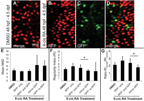
Two-dimensional pattern analysis of LWS cones in LWS:PAC(H) embryos exposed to RA: Retinoic acid induced LWS1-expressing cones do not disrupt the LWS2 cone mosaic. Regions from confocal images at 60X magnification obtained from whole mounted LWS:PAC(H) eyes treated with DMSO or 0.3 µM 9-cis RA 48 hpf to 4.5 dpf were used for pattern analysis. A-D. Representative regions used to determine the mean nearest neighbor distance (NND) and Regularity Index (RI) of LWS cones in control and 9-cis RA-treated eyes. Regions from DMSO treated retinas expressed only LWS2:RFP cones (red) (A), while regions from 9-cis RA treated retinas (B-D) contained RFP+ (red), GFP+ (green), and RFP+/GFP+ (yellow) cones. B-D. Images showing RFP signal only (B), GFP signal only (C), and merge of both signals (D) in regions of 9-cis RA treated retina. Graphs of NND (E, left three bars) and RI (F, left three bars) corresponding to regions from DMSO treated (DMSO) and 9-cis RA treated retinas. 9-cis RA treated retinas were further divided into groups: regions of retina where only RFP+ cones are present (RFP+ only), and regions containing both RFP+ and GFP+ cones (RFP+ and GFP+) when all labeled cells (regardless of RFP or GFP expression) are treated as the same cell type. Graphs also indicate NND (E, right two bars) and RI (F, right two bars) for 9-cis RA treated retinas when GFP+ cones are subtracted from the RFP+ and GFP+ mosaic (minus GFP+) and when the pattern of GFP+ cones to RFP+ cones (GFP+ to RFP+) is analyzed. G. Graph showing the ratio of RI from each indicated region (RIsample) with that of the average RI from 1000 generated random samples of the same number of cells (RIrandom). The asterisk (*) in F and G indicates p<0.05 for a two-tailed Student’s t-test between the indicated groups. Error bars represent standard deviation (n = 7 regions from 4 eyes, DMSO; 6 regions of RFP+ only and 10 regions RFP+ and GFP+ from 6 eyes, RA). |
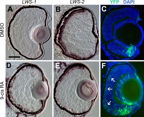
Spatial relationship of RA signaling activity compared with expression of LWS1 and LWS2. A-C. Sections obtained from a RARE:YFP transgenic embryo treated with DMSO 48 hpf-100 hpf, then labeled for LWS1 mRNA expression (A), LWS2 mRNA expression (B), or YFP immunofluorescence (C; counterstained with DAPI). D-F. Sections obtained from a RARE:YFP transgenic embryo treated with 0.3 µM 9-cis RA 48 hpf– 100 hpf, then labeled for LWS1 mRNA expression (D), LWS2 mRNA expression (E), or YFP immunofluorescence (F). Expanded RA signaling domain (arrows) is similar to the LWS1 expression domain. Scale bar in A (applies to all) = 50 µm. |

An “LWS Transition Zone” exists in ventral retina of juvenile fish. Sections of retina from one-month old juvenile retinas are shown. A and B. Two different sections from separate LWS:PAC(H) transgenic fish showing LWS1:GFP+ cones (green fluorescence; long arrows), LWS2:RFP+ cones (red fluorescence), and cones co-expressing LWS1:GFP and LWS2:RFP (yellow, short arrows). B. The asterisk (*) indicates a cone expressing only LWS2:RFP at the ventral periphery. C. Dual in situ hybridization using cDNA probes for LWS1 (purple) and LWS2 (red/pink). Cones expressing LWS1 only are indicated by long arrows; cones co-labeled with both probes are indicted by short arrows. The asterisk (*) indicates an LWS2 singly labelled cone at the ventral periphery. Scale bar in A (applies to all) = 25 µm; v, ventral; d, dorsal. |

RA signaling continues in juvenile retinas and the signaling domain includes red opsin+, LWS1+ cones. Sections of retina from one-month old fish are shown, with ventral pole at bottom of images. A. Immunocytochemistry to reveal YFP signal and DAPI in a section from RARE:YFP retina. Arrows indicate YFP signal in the outer nuclear layer, corresponding to the location of cone photoreceptors. B. In situ hybridization for topaz-YFP mRNA in a section from a RARE:YFP retina. C and D. LWS:PAC(H) were crossed with RARE:YFP and doubly transgenic juvenile retina sections were examined for RFP, GFP, and YFP fluorescence. Although YFP and GFP cannot be fully resolved, the YFP+ signaling domain coincides with the GFP (LWS1) expression domain. The GFP expression domain (reporting LWS1) is designated by dashed arc (C and D), while the YFP domain (reporting RA signaling) is between the dashed straight lines (D). E. Sections from RARE:YFP juvenile retinas were stained with antibodies for YFP, ZPR1 (labels red- and green-sensitive cones), and DAPI; merged image is shown. Colabel of YFP and ZPR1 is yellow. Arrows indicate ZPR1+ cones that are YFP+. F. Sections from RARE:YFP juvenile retinas were stained with antibodies for YFP, red opsin (pan-LWS), and DAPI; merged image is shown. Arrows indicate red opsin+ cone outer segments that are continuous with YFP+ cell bodies. G. Dual in situ hybridization for topaz-YFP (purple) and LWS1 (red/pink) mRNA in a section from one-month old RARE:YFP retina. Arrows indicate examples of some of the colabeled cones. Scale bar in A = 25 µm; scale bar in C (applies to B-G) = 25 µm; v, ventral; d, dorsal. |