
- Title
-
Hsp47 mediates Cx43-dependent skeletal growth and patterning in the regenerating fin
- Authors
- Bhadra, J., Iovine, M.K.
- Source
- Full text @ Mech. Dev.

serpinh1b is differentially expressed in wild-type, sof b123 and alf dty86. Whole mount in situ hybridization shows increased expression in alf dty86 (C) and decreased expression in sof b123 (B) compared to wild-type (A). In situ hybridization on cryosection of a WT-5 dpa fin (D) reveals localization of serpinh1b in the blastema (b), basal layer of epidermis (ble) and skeletal precursor cells (*). Epidermis is denoted by ‘e’, mesenchyme by ‘m’, lepidotrichia by ‘l’ and actinotrichia by ‘a’. The amputation plane is indicated in panels A and B. The amputation plane is beyond the field of view in C. The scale bars in C and D are 50µm; the scale bar in C applies to the images in A, B, and C. |

ZFIN is incorporating published figure images and captions as part of an ongoing project. Figures from some publications have not yet been curated, or are not available for display because of copyright restrictions. |
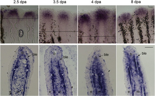
serpinh1b expression over time in regenerating fins. Whole mount in situ hybridization for serpinh1b at different time points on regenerating fins (top). The amputation plane is indicated in 2.5, 3.5 and 4 dpa fins. For 8 dpa fins, the amputation plane is outside the field of view. In situ hybridization on cryosectioned fins at different time points (bottom). Similar localization of serpinh1b gene in basal layer of epithelium (ble) and skeletal precursor cells (*) is observed. Blastema (b), basal layer of epidermis (ble), and skeletal precursor cells (*). The scale bars are 50µm. The scale bar in the upper 8 dpa panel applies to all panels for whole mount images. The scale bar in the lower 8 dpa panel applies to all panels for cryosections. EXPRESSION / LABELING:
|

Morpholino mediated knockdown of Hsp47 results in reduced expression in WT regenerating fins. (A) Longitudinal sections of WT 5 dpa fins following treatment with standard control or targeting MO against serpinh1b. Hsp47-MO treated fins show reduced level of Hsp47 expression. Hsp47 (red), nuclei are stained with the far red dye To-Pro (blue). ‘ble’ indicates basal layer of epithelium; ‘b’ blastema; ‘m’ mesenchyme, and ‘e’ epidermis. Scale bar is 10µm, and applies to all panels in A. (B) Immunoblots confirming reduced level of Hsp47 following Hsp47 knockdown. Hsp47 MO treated fins (MO) are compared to fins injected with standard control MO. Tubulin is used as loading control. (C) Bar graph depicting the ImageJ analysis of the ratio of Hsp47 levels between sof b123 or alf dty86 and wild-type (normalized by the tubulin loading control). Hsp47 protein levels are reduced in sof b123 and upregulated in alf dty86 relative to WT (represented as a relative density of 1). |
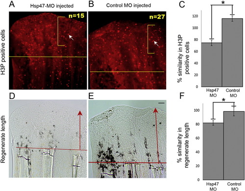
Morpholino mediated knockdown of Hsp47 causes reduced cell proliferation and fin length. All fins were amputated at the 50% level prior to injection and electroporation. Fin rays treated with targeting or control MO (injected) were measured and compared to their uninjected sides. The ratio of the injected side (targeting MO or control MO) and the control uninjected side, multiplied by 100, is the percent similarity. Percent similarity of greater than 100% reflects the fact that the experimental side can be measurably larger than the control uninjected side. (A–C) Total number of H3P positive cells (white arrows identify one positive cell in each panel) were counted in injected and uninjected sides for Hsp47-MO (A) and Hsp47-5MM (B) treated fins. (C) The bar graph shows reduced level of H3P positive cells in Hsp47 MO injected fins compared to the control MO. (D–F) Total regenerate length in Hsp47-MO (D) and Hsp47-5MM (E) injected fins was measured by evaluating the distance between the distal tip of the fin and the amputation plane (indicated by red arrows) and normalized to the respective uninjected side. (F) Bar graph shows reduced regenerate length in Hsp47 MO treated fins. Scale bar is 50µm and applies to all panels. The student′s t-test was performed (p < 0.05) to determine significance. PHENOTYPE:
|

Morpholino mediated knockdown of Hsp47 causes reduced segment length and a small change in bone regenerate length. Calcein staining was used to measure segment length and bone regenerate length in the regenerate. The amputation plane is indicated by a white line. Fin rays treated with targeting or control MO (injected) were measured and compared to their uninjected sides. The ratio of the injected side (targeting MO or control MO) and the control uninjected side, multiplied by 100, is the percent similarity. Percent similarity of greater than 100% reflects the fact that the experimental side can be measurably larger than the control uninjected side. (A) Segment length in Hsp47-MO and Hsp47-5MM injected fins was measured as the distance between the 1st and the 2nd joints (indicated by white arrow heads) distal to the amputation plane. Bone regenerate length was measured as the ratio of the distance of the regenerated length of the calcein stained bone matrix (marked as a) and the total regenerate length (marked as b). The treatment side was normalized against the uninjected side. (B) Bar graph shows reduced segment length following Hsp47 MO injection (p < 0.05). (C) Bar graph shows small but significant reduction in bone regenerate length. The scale bar is 50µm and applies to both panels in A. The student′s t-test was performed (p < 0.05) to determine significance. PHENOTYPE:
|

The short segment phenotype following Hsp47-MO treatment is due to formation of premature joints. ZNS5 immunostaining detects osteoblasts and joint-forming cells. Brightfield (left) and ZNS5 immunostaining (right) images are shown. ZNS5 positive joints (indicated by arrows) are observed in both standard control (top row) and targeting MO (bottom row) treated fins. Scale bar is 50µm and applies to all panels. |
|
ZFIN is incorporating published figure images and captions as part of an ongoing project. Figures from some publications have not yet been curated, or are not available for display because of copyright restrictions. |

Localization of Hsp47 and Collagen type II in WT-5 dpa fins. Confocal images of a WT-5 dpa longitudinal fin section immunostained with Hsp47 (red) and collagen type II (green) and counterstained with To-pro to detect nuclei (blue). Arrows indicate actinotrichia. ‘ble’ is basal layer of epithelium, ‘e’ epidermis and ‘m’ mesenchyme. Scale bar is 10µm and applies to all panels. EXPRESSION / LABELING:
|
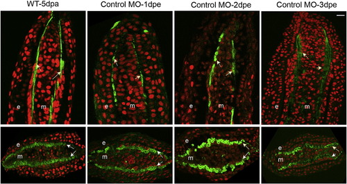
Examination of actinotrichia in fin sections following injection with standard control MO at different timepoints. Single plane confocal sections were obtained for longitudinal and transverse sections immunostained with Collagen type II (green) and counterstained with propidium iodide (red). Untreated fins are labeled as ‘WT-5 dpa’. Longitudinal (top) and transverse (bottom) sections show regeneration of actinotrichia at different time points following injection and electroporation with standard control MO. Arrows indicate actinotrichia, ‘e’ is epidermis and ‘m’ mesenchyme. Scale bar is 10µm and applies to all panels. EXPRESSION / LABELING:
|
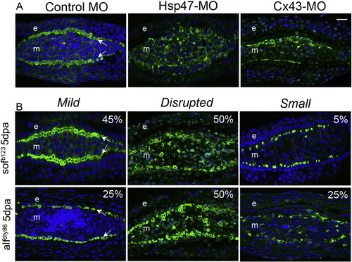
Disruption of actinotrichia following changes in Hsp47 or Cx43 expression. Single plane confocal sections are obtained for transverse sections immunostained with Collagen type II (green) and counterstained for nuclei with To-pro (blue). (A) Fins are injected with standard control MO, Hsp47-MO or Cx43-MO and evaluated at 1 dpe. Col II expression is disrupted following Hsp47 and Cx43 knockdown. (B) Variable actinotrichia phenotypes observed in 5 dpa transverse sections of both sof b123 and alf dty86. The defects were categorized as mild, disrupted organization, or small. Arrows indicate actinotrichia, ‘e’ is epidermis and ‘m’ mesenchyme. Scale bar is 10µm and applies to all panels. |

serpinh1b expression level is upregulated during regeneration. serpinh1b expression is undetectable during ontogeny. Expression of serpinh1b at 5 dpa is shown for comparison. The expression level is reduced at 14 dpa. The amputation plane is indicated in 5 dpa fin. The amputation plane is beyond the field of view in 14 dpa fin. Scale bar indicates 50µm and applies to all panels. |
Reprinted from Mechanisms of Development, 138 Pt 3, Bhadra, J., Iovine, M.K., Hsp47 mediates Cx43-dependent skeletal growth and patterning in the regenerating fin, 364-74, Copyright (2015) with permission from Elsevier. Full text @ Mech. Dev.