
- Title
-
Hypoxia-Induced Retinal Neovascularization in Zebrafish Embryos: A Potential Model of Retinopathy of Prematurity
- Authors
- Wu, Y.C., Chang, C.Y., Kao, A., Hsi, B., Lee, S.H., Chen, Y.H., Wang, I.J.
- Source
- Full text @ PLoS One
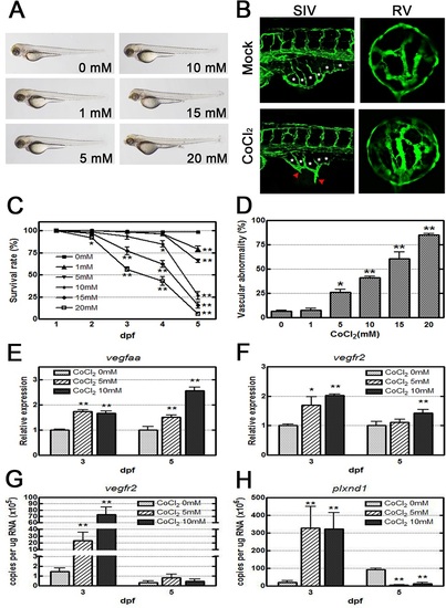
CoCl2 induces abnormal neovascularization in zebrafish embryos. (A) Morphological images obtained by an optical microscope revealed no severe phenotype in CoCl2-treated Tg(fli1a:EGFP) embryos at 3 dpf. (B) With fluorescence excitation, ectopic SIV and excessive retinal vascularization are shown in CoCl2-treated embryos compared with the untreated control. (C, D) A dose-dependent decrease in survival rates (four independent experiments; n = 35 in each group) in embryos treated with increasing concentrations (0–20 mM) and an increase in the vascular defect occurrence rate in the SIV and retinal vessels are shown (three independent experiments; n = 35 in each group). (E, F) Real-time RT-PCR data show that CoCl2 treatment causes overexpression of vegfaa and vegfr2 mRNAs in zebrafish. (G,H) Absolute quantification of the copy number of vegfr2 and plxnd1 by real-time PCR are reduced at 5 dpf versus 3 dpf. Each bar represents the mean ± SEM. * (p < 0.01) and ** (p < 0.001) compared with the mock control group. SIV, subintestinal vessel; RV, retinal vessel. EXPRESSION / LABELING:
PHENOTYPE:
|

Effects of CoCl2 and the VEGF inducer GS4012 on retinal neovascularization. (A, B) Time-course and dose-dependent effects of GS4012 on the survival rate and the vascular defect-occurrence rates of Tg(fli1a:EGFP) embryos are shown. Embryos were treated with 2.5, 5, or 7.5 µg/mL of GS4012 and 5 mM CoCl2 for 1, 2, 3, 4, and 5 dpf. Each bar of the survival rate and the vascular defect-occurrence rates represents the mean ± SEM (n = 50 in each group). * p < 0.01 and + p < 0.001, as compared with the control group and the equivalent concentration of GS4012 group, respectively. (C–J) Fluorescence microscope observations of the retinal vessels of treated Tg(fli1a:EGFP) embryos are shown. At 3 dpf, compared with the untreated control (C), both CoCl2 and GS4012 induced vessel branching in the retina (D, E). Zebrafish embryos cotreated with CoCl2 and GS4012 showed severe branching and disorganization in the retinal vasculature (F). At 5 dpf, compared with the untreated control (G), CoCl2-treated retinal vessels were narrow, indicating vasoconstriction (H), and GS4012-treated vessels appeared tortuous and twisted (I). Furthermore, cotreatment with CoCl2 and GS4012 induced a complex, highly disorganized, and tortuous vasculature (J). (K) The left panel shows the method for counting and measuring branch points (red asterisks) and vessel diameters (yellow line). The vessel diameters were measured using Image J (three randomly chosen positions). These data consist of observed characteristics. Each bar of the branch points and vessel diameter chart represents the mean ± S.D. (n = 5 in each group). * p < 0.01, as compared with the control group. + p < 0.01 and ++ p < 0.001, as cotreatment groups compared with the equivalent concentration of GS4012 and CoCl2 group, respectively. EXPRESSION / LABELING:
PHENOTYPE:
|
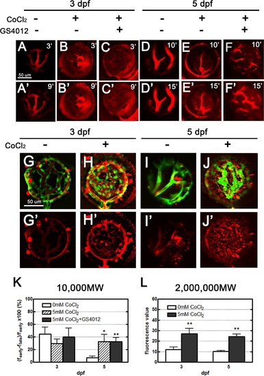
Leakage analyses of CoCl2-treated retinal vasculature performed using two types of fluorescent dyes: 10,000 MW dextran, and 2,000,000 MW TAMRA. (A–F′) Injected dextran (red) in the vessels became obscured, with apparent dextran leakage within 9 and 15 minutes in treated zebrafish embryos 3 and 5 dpf, respectively (B′, C′, E′, F′); however, in the control embryos, leakage was observed in the retinal vessels (A′). No leakage was apparent in the normal tight endothelium of 5-dpf embryos (D′). (G–J′) Upon injection, most of the TAMRA dye (red) was contained in the vasculature, and leakage was observed 24 hours later. Scant leakage occurred at the center of the control retina (G, I), whereas apparent leakage was observed in CoCl2-treated vasculatures (H, J). (K, L) The fluorescence values in 3- and 5-dpf embryos were quantified using Image J to show the dynamic changes of the 10,000 MW and 2,000,000 MW dyes. Data are presented as the mean ± standard deviation from three to five embryos. EXPRESSION / LABELING:
PHENOTYPE:
|

VEGF inhibitors rescue overangiogenesis and leakage in the CoCl2-induced hypoxic retinal vasculature. (A) Three conditions were designed to mimic the clinical situation and to examine the effects of treatment. Method I: Tg(fli1a:EGFP) embryos were treated with CoCl2 (black line) and the inhibitor (red dotted line) simultaneously from 1 dpf to 5 dpf. Method II: Prior to CoCl2 treatment (black line) at 3 dpf, we immersed embryos in the inhibitor (red dotted line) for 2 days. Method III: Following CoCl2treatment, the inhibitor was added to the solution at 3 dpf. All embryos were injected with TAMRA dye at 4 dpf, and their retinal vasculatures were subsequently observed at 5 dpf. (B) CoCl2-treated embryos showed TAMRA dye seepage (red) from the intraocular vessels (green). Similar leakages and excessive neovascularization were appeared in the control IgG-treated groups (CoCl2 and normal human control IgG cotreatment). Three candidate inhibitors, SU5416, bevacizumab, and ranibizumab, reversed the effect of CoCl2 on retinal vessels. (C) Statistical analyses of branch points showed significant rescue in inhibitor-treated embryos. The total numbers of embryos for analyses are indicated on the top of each bar. (D) However, the survival rates (four independent experiments, n = 35 in each) showed low toxicity under the Method I condition. Each bar represents the mean ± SEM of * (p < 0.0125) compared with the CoCl2 = 5 mM group. EXPRESSION / LABELING:
|

Early development of retinal neovascularization in zebrafish. (A–C) Confocol images of GFP expression in the eye of Tg(fli1a:EGFP) zebrafish. (A) DAPI staining (blue) was used to orient the ocular blood vessels at 6 dpf. (B) In the surface vasculature, blood enters through the nasal vessel (nrv) and exits through the dorsal (drv) and ventral (vrv) vessels. Blood from the retinal vessels flows through the annular collection duct (asterisks) into the surface vessels. (C) Lateral view of the retinal vessel network. The retinal artery or its presumptive primordium is indicated by the red arrowhead. (D) Retinal vessels at 6 dpf. (E) At 1 dpf, uniform growth of the retinal vessels from the central retinal artery was observed. (F) The vessels assumed a cup shape at 2 dpf. (G) The retinal vessels increased in number at 3 dpf. (H) Vessel branching was apparent and the vascular architecture complexity increased in the retina 4 dpf. (I) Vessel branching and organization further developed, as indicated by the formation of numerous branch points and sprouts at 5 dpf. |

Effect of SU5416 on retinal vessels at 3 dpf. (A) SU5416 inhibited the development of the retinal vessels. (B) SU5416 reversed the effect of CoCl2 on the retinal vessels and prevented the leakage of TAMRA dye through the vessels. |