
- Title
-
A transgenic zebrafish model for monitoring xbp1 splicing and endoplasmic reticulum stress in vivo
- Authors
- Li, J., Chen, Z., Colorni, A., Ucko, M., Fang, S., Du, S.J.
- Source
- Full text @ Mech. Dev.

Construction of the Tg(ef1α:xbp1δ-gfp) transgene and expression of XBP1Δ-GFP in live zebrafish embryos. (A) A partial zebrafish xbp1 cDNA sequence (239 bp) containing the 26 nt IRE1 target sequence was cloned upstream of the EGFP coding sequence. A myc-tag sequence was also included in frame at 5′ end of the xbp1 sequence. Under normal conditions without ER stress, the xbp1δ-gfp transcripts are not spliced and protein translation stops prior to the GFP coding sequence. Upon ER stress, the 26 nt IRE1 target sequence is spliced out from the xbp1δ-gfp mRNA transcripts, leading to a frame shift and the expression of the XBP1Δ-GFP fusion protein. (B) Expression of the XBP1Δ-GFP in live zebrafish embryos. XBP1Δ-GFP expression was directly observed in the F1 transgenic zebrafish embryos from one cell stage to 24 hpf. A strong XBP1Δ-GFP expression was observed in transgenic embryos with the transgene derived maternally. In contrast, transgenic embryos with paternally derived transgene showed no XBP1Δ-GFP expression from one cell stage to 12 hpf. A weak expression was detected at 24hpf. Non-transgenic fish embryos were used as control. |

Maternal expression of XBP1Δ-GFP and the spliced isoform of xbp1 transcripts. (A, B) Direct observation of XBP1Δ-GFP expression in unfertilized zebrafish oocytes dissected from Tg(ef1α:xbp1δ-gfp) transgenic (A) or non-transgenic (B) females. (C) RT-PCR analysis of endogenous xbp1 mRNA splicing and BIP expression in unfertilized eggs, testis from adult fish, and embryos at 3 hpf and 24 hpf. |

XBP1Δ-GFP expression in homozygous transgenic zebrafish larvae. (A and B) XBP1Δ-GFP expression in live Tg(ef1a:xbp1δ-gfp) transgenic zebrafish larvae at 24 hpf. (C) Non-transgenic fish larvae at 24 hpf with GFP filter. (D) Bright filed of Tg(ef1a:xbp1δ-gfp) transgenic zebrafish larvae at 24 hpf. (E and G) XBP1Δ-GFP expression in Tg(ef1a:xbp1δ-gfp) transgenic zebrafish larvae at 72 (C) or 150 (G) hpf. (F and H) Non-transgenic fish larvae at 72 (F) or 150 (H) hpf. (I and J) Expression and splicing of the Tg(ef1α:xbp1δ-gfp) transgene (I) or the elongation factor 1α (J) at day 3, 5 and 7. |

Characterization of DTT and tunicamycin induced ER stress in Tg(ef1α:xbp1δ-gfp) transgenic zebrafish embryos. Transgenic embryos with the paternally derived Tg(ef1α:xbp1δ-gfp) transgene were treated with 0.5 mM of DTT, 1 µg/ml or 2 µg/ml of tunicamycin between 24 and 48 hpf. XBP1Δ-GFP expression and mRNA splicing were analyzed at 48 hpf after the DTT or tunicamycin treatment. (A, E) Side and dorsal views of non-transgenic fish embryos at 48 hpf. (B, F) Side and dorsal views of Tg(ef1α:xbp1δ-gfp) transgenic embryos at 48 hpf without DTT treatment. (C, G) XBP1Δ-GFP expression in transgenic embryos at 48 hpf with 24 h of 0.5 mM DTT treatment. (D, H) XBP1Δ-GFP expression in transgenic embryos at 48 hpf with 24 h of 1 µg/ml tunicamycin treatment (D) or without the treatment (H). (I) RT-PCR analysis shows the expression and splicing of the Tg(ef1α:xbp1δ-gfp) transgene and the endogenous xbp1 gene in DTT treated or control paternal transgenic (P-) and wild type (WT-) embryos. P-Endo represents xbp1 expression from the endogenous gene. P-Exo represents xbp1 expression from the exogenous Tg(ef1α:xbp1δ-gfp) transgene. WT-Endo represents xbp1 expression from the endogenous gene in wild type embryos. Arrows indicate spliced xbp1 transcripts. (J). Western blot analysis shows the expression of the myc-tagged XBP1Δ-GFP fusion protein derived from the paternal transgenic line at 48 hpf in control and DTT treated embryos. (K). RT-PCR analysis shows the splicing of endogenous and exogenous xbp1 in paternal transgenic zebrafish embryos treated with 1 µg/ml (TUN-1) and 2 µg/ml (TUN-2) tunicamycin for 24 hpf between 24 and 48 hpf. EF1α expression was analyzed as an internal control. Arrows indicate spliced xbp1 transcripts. (L). RT-PCR analysis shows the expression of ER stress markers, xbp1 and BIP, in zebrafish embryos treated with 0.5 mM DTT for 24 hpf between 24 and 48 hpf. |
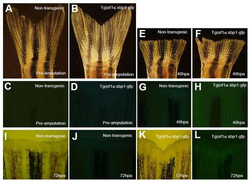
Analysis of Tg(ef1α:xbp1δ-gfp) expression during caudal fin regeneration. Non-transgenic and Tg(ef1α:xbp1δ-gfp) transgenic fish (4 months) were subjected to caudal fin amputation. XBP1Δ-GFP expression were analyzed at preamputation and 48 and 72 hours post amputation (hpa). A–D. Bright field (A, B) and whole mount fluorescent (C, D) images of caudal fins of non-transgenic and Tg(ef1α:xbp1δ-gfp) transgenic fish at preamputation. E–H. Bright field (E, F) and whole mount fluorescent (G, H) images of caudal fins of non-transgenic and Tg(ef1α:xbp1δ-gfp) transgenic fish at 48 hpa. I–L. Bright field (I, K) and whole mount fluorescent (J, L) images of caudal fins of non-transgenic and Tg(ef1α:xbp1δ-gfp) transgenic fish at 72 hpa. |
Reprinted from Mechanisms of Development, 137, Li, J., Chen, Z., Colorni, A., Ucko, M., Fang, S., Du, S.J., A transgenic zebrafish model for monitoring xbp1 splicing and endoplasmic reticulum stress in vivo, 33-44, Copyright (2015) with permission from Elsevier. Full text @ Mech. Dev.