
- Title
-
Sox7 controls arterial specification in conjunction with hey2 and efnb2 function
- Authors
- Hermkens, D.M., van Impel, A., Urasaki, A., Bussmann, J., Duckers, H.J., Schulte-Merker, S.
- Source
- Full text @ Development

Disrupted blood circulation in sox7 zebrafish mutants. (A) sox7 in situ hybridization of 3dpf wild-type embryos (left panel, lateral view; right panel, dorsal view) showing sox7 expression in all main vessels. (B) Schematic diagram of the sox7hu5626 allele with a premature stop-codon after amino acid 53 (red asterisk). HMG, high mobility group box; TAD, trans-activating domain. (C) Overall normal appearance of sox7hu5626 heterozygous sibling and homozygous mutant at 2dpf. (D) On average, 59% of sox7hu5626 mutants display disturbed blood flow at 2.5dpf. Percentages of pooled embryos from four independent experiments (total of 275 embryos). Percentages can vary substantially between different backgrounds. (E) kdrl:GFP;gata1:dsRed-positive sox7hu5626 mutants lack functional blood circulation in the trunk, whereas heterozygous siblings have normal circulation at 2.5dpf. Right panels: higher magnifications of boxed area. EXPRESSION / LABELING:
PHENOTYPE:
|
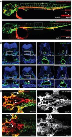
sox7 mutants show an altered morphology of the LDA while displaying normal DA-PCV segregation in the trunk. (A) Rhodamine-dextran angiograms (injected into the PCV at the position of the arrow) of sox7hu5626 fli1a:eGFP siblings (upper panel) and mutants (lower panel) at 2dpf. Insets depict the rhodamine-dextran channel of the boxed area with higher magnification highlighting the lack of dye uptake in the DA of sox7hu5626 mutants. (B) Transverse sections of fli1a:eGFP-positive sox7hu5626 sibling and mutant embryos at 2dpf, stained with anti-GFP (green) and DAPI (blue). In sox7hu5626 mutants, the aortic morphology is disturbed at the position of LDA/DA fusion (arrows) while being unaffected more anteriorly and posteriorly to this position. For relative positions of sections, see supplementary material Fig. S3. (C) Dorsal view of sox7hu5626 kdrl:mCherry;flt4:mCitrine sibling and mutant embryos at 2.5dpf. Right panel kdrl:mCherry channel only. Shunt formation (arrow) occurs in sox7hu5626 mutants at the position of LDA fusion. (L) DA, (lateral) dorsal aorta; PCV, posterior cardinal vein; CCV, common cardinal vein. |

sox7 mutants develop a circulatory short-loop phenotype. (A) sox7hu5626 kdrl:eGFP;gata1:dsRed mutants at 2.5dpf with shunt formation resulting in a circulatory short-loop (middle and right panel) in contrast to normal circulation in sox7hu5626 siblings (left panel). Middle panel depicts ectopic connection between LDA and PHS, right panel between LDA and CCV. Lower panel depicts only gata1:dsRed with detailed schematic representation of blood flow. CCV, common cardinal vein; LDA, lateral dorsal aorta; PHS, primary head sinus; VA, ventral aorta. (B) Schematic representation of normal blood flow in wild-type embryos compared with the short-circuit in sox7hu5626 mutants at 2.5dpf. Dashed circles highlight the position of the heart. (A,B) Lines depict blood flow in arteries (red) and veins (blue). (C) Stills of time-lapse movies of kdrl:eGFP-positive sox7 mutant and sibling at the time point of ectopic connection formation (see supplementary material Movies 3 and 4). ECs of the LDA (outlined by red lines) connect to ECs of the CCV (outlined by blue lines) in the sox7 mutant at 26hpf (yellow arrow). (D) Quantification of the phenotypes in 3dpf sox7hu5626;sox18hu10320 loss-of-function embryos. All sox7hu5626;sox18hu10320 double homozygous mutants have a short-loop of circulation and shunts between DA and PCV, whereas sox7/sox18+/+ and sox7/sox18+/ embryos have no shunts between DA and PCV. The remaining, non-shown genotypic combinations of sox7;sox18 embryos all have 100% wild-type phenotype. Bars show percentages of embryos of a representative experiment (total of 77 embryos). (E) Hematoxylin and Eosin stained transverse (left panel) and sagittal (right panel) cross-sections of trunks of 2dpf sox7hu5626 siblings (top) and homozygous mutants (bottom) showing normal DA-PCV segregation in both siblings and mutants (arrows). PHENOTYPE:
|
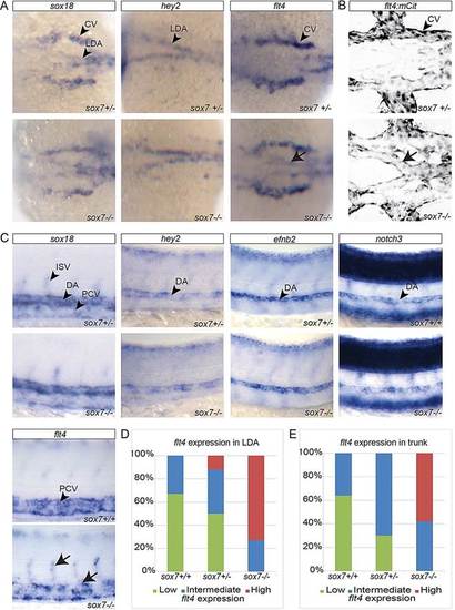
flt4 expression is altered in arterial cells of sox7 mutants. (A-C) sox7hu5626 siblings (upper panel) and mutants (lower panel). (A) In situ hybridization for sox18, hey2 and flt4 in sox7hu5626 heterozygous and mutant embryos at 20hpf (dorsal view). sox7hu5626 mutants display elevated flt4 expression levels in the LDA (arrow). (B) Dorsal view of flt4:mCitrine (flt4:mCit)-positive sox7hu5626 heterozygous and homozygous mutant at 26hpf showing elevated mCitrine expression in the LDA of sox7hu5626 mutant (arrow) compared with sox7hu5626 siblings. (C) Lateral view of the trunk region of sox7hu5626 siblings and mutants with in situ hybridization for sox18, hey2, efnb2, notch3 and flt4 at 24-27hpf. sox7hu5626 mutants display higher flt4 expression in DA and ISVs (arrows) compared with siblings. (D) Quantification of 20-24hpf embryos based on their flt4 expression levels (in situ hybridization) in the LDA. Seventy-three percent of sox7hu5626 mutants have high expression of flt4 in LDA region at 20hpf (total of 30 embryos). (E) Quantification of 24-27hpf embryos based on their flt4 expression levels (in situ hybridization) in the trunk. Fifty-eight percent of sox7hu5626 mutants have elevated flt4 expression, which was never observed in siblings (total of 57 embryos). (L)DA, (lateral) dorsal aorta; (P)CV, (posterior) cardinal vein; ISV, intersegmental vessel. EXPRESSION / LABELING:
|

efnb2a;efnb2b double mutants develop a circulatory short-circuit phenotype and sox7 genetically interacts with efnb2a. (A) Schematic diagram of efnb2ahu3393 and efnb2bhu2971alleles with premature stop codons (red asterisks) at amino acids 86 and 78, respectively. Eph, ephrin domain (green), Cd, cupredoxin domain (blue). (B) Quantification of efnb2ahu3393 and efnb2bhu2971 single and double mutant phenotypes revealed circulatory short-looping in 11% of efnb2ahu3393/;efnb2bhu2971+/ embryos and a significant increase of short-looping in 42% of double homozygous mutants (Student′s t-test, P=0.003 compared with efnb2ahu3393/;efnb2bhu2971+/). The genotype combinations not shown all displayed 100% wild-type phenotype. Bars represent combined results of five independent experiments (total of 128 embryos). (C) Blood circulation in gata1:dsRed-positive efnb2ahu3393;efnb2bhu2971 single and double mutant embryos at 2.5dpf. Note normal circulation in single mutants (upper panel) and short-loop phenotype in efnb2ahu3393;efnb2hu2971 double mutants (lower panel). Schematic representation of blood flow in double mutants (lower right panel). Lines depict blood flow in arteries (red) and veins (blue). VA, ventral aorta; PHS, primary head sinus; CCV, common cardinal vein. (D) Combinations of sox7hu5626, efnb2ahu3393 and efnb2bhu2971 loss-of-function alleles result in increasingly severe effects on the circulatory system. All sox7hu5626;efnb2ahu3393 double homozygous mutant embryos exhibit a significant (Student′s t-test, P=0.002) increase in the penetrance of the short-circuit phenotype compared with the only partially penetrant sox7hu5626 single mutants or the efnb2ahu3393 single mutants lacking the short-loop defect completely. The genotype combinations not shown all displayed 100% wild-type phenotype. Bars represent combined results of five independent experiments (total of 507 embryos). (B,D) Embryos were analyzed at 2.5dpf. Phenotypes classified as ‘others’ are edema, total lack of circulation and/or shunts in trunk region. EXPRESSION / LABELING:
PHENOTYPE:
|
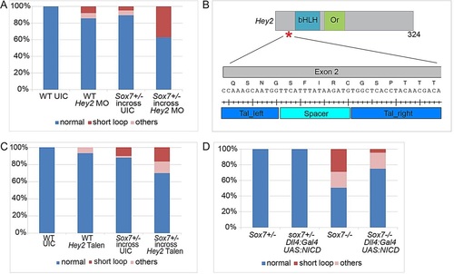
sox7 synergizes with hey2 and functions upstream of Notch ICD in LDA development. (A) The partially penetrant short-loop phenotype of both sox7hu5626 mutants (n=104) and hey2 morphants (0.3ng, n=63) is exacerbated when the hey2 MO is injected into a sox7hu5626+/- in-cross (n=35). Bars show the percentage of embryos from a representative experiment. (B) Schematic diagram of Hey2, indicating the TALEN target site in exon 2, which is upstream of the coding region of the basic helix-loop-helix (bHLH) DNA-binding domain and the orange (Or) domain. (C) Transient injections of 10pg per embryo hey2 TALEN mRNA into wild-type and sox7hu5626 heterozygous in-cross. There is a moderate enhancement of penetrance of the short-loop phenotype in sox7hu5626 in-cross injected with hey2 TALEN (n=140 embryos) compared with un-injected sox7hu5626 in-cross (n=100 embryos) and hey2 TALEN injections into wild-type embryos (n=90 embryos). (D) Arterial specific UAS:NICD overexpression using the dll4:Gal4 driver line significantly (Student′s t-test, P=0.004) rescues the short-loop circulatory phenotype in sox7 mutants: short-loop phenotype in 27% of mutants without NICD overexpression (n=69 embryos) and in 3% with NICD overexpression (n=44 embryos). Bars represent pooled embryos of three independent experiments (total of 300 embryos). (A,C,D) Embryos were analyzed at 2.5dpf. Phenotypes classified as ‘others’ are edema, total lack of circulation and/or shunts in trunk region. PHENOTYPE:
|