
- Title
-
IFT46 plays an essential role in cilia development
- Authors
- Lee, M.S., Hwang, K.S., Oh, H.W., Ji-Ae, K., Kim, H.T., Cho, H.S., Lee, J.J., Yeong Ko, J., Choi, J.H., Jeong, Y.M., You, K.H., Kim, J., Park, D.S., Nam, K.H., Aizawa, S., Kiyonari, H., Shioi, G., Park, J.H., Zhou, W., Kim, N.S., Kim, C.H.
- Source
- Full text @ Dev. Biol.
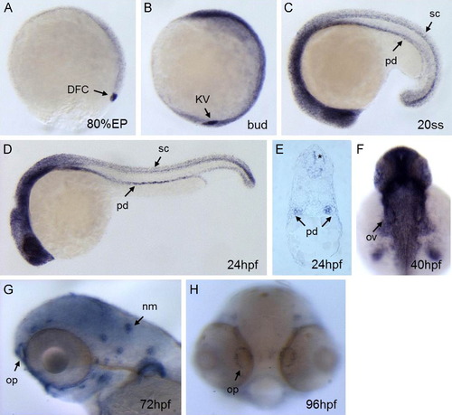
Expression pattern of ift46 in zebrafish embryos. (A) Whole-mount in situ hybridization shows ift46 expression at 80% epiboly in the dorsal forerunner cells (DFC, arrow). (B) ift46 is strongly expressed in Kupfferós vesicle (KV, arrow) at bud stage, (C, D) At 20-somite stage and 24 hpf ift46 expression is detected in pronephric ducts (pd), eyes, ears, spinal cord (sc) and diffusely in the brain. (E) Cross-section showing ift46 expression in the pronephric ducts (arrow) and spinal cord (asterisk). (F) At 40 hpf, ift46 is widely expressed in brain, eyes, otic vesicles (arrow) and pectoral fins. (G, H) At 72 and 96 hpf, ift46 is expressed in the olfactory pits (op) and neuromast (nm) hair cells (arrow). EXPRESSION / LABELING:
|

ift46 morphants develop phenotypes associated with ciliary dysfunction. (A) Wild-type embryos show normal morphology. (B) ift46 morphants are characterized by curved body axis, the development of pronephric cyst and pericardiac edema at 96 hpf. (C–D) Cross-sections of wild-type zebrafish at the glomerular–tubular region of pronephros at 72 hpf show normal morphology of pronephric glomeruli and ducts. (Có–Dó) ift46 morphant embryos display a grossly distended cyst (asterisk) in place of glomeruli (gl) and dilated pronephric ducts (pd) (arrow). Scale bar, 50 µm. (E–F′) Histological sections of the eyes of wild-type and ift46 morphants at 5 dpf. The retina in wild-type control (E and E′) has normal laminated structures, including retinal pigment epithelium (RPE), outer segment (OS), outer plexiform layer (OPL) and inner nuclear layer (INL). (E) and (F) scale bar, 200 µm. The outer segment (OS) (bracket) is thinner in ift46 morphants (F′) compared to the control (E′). Scale bar, 100 µm. (G–J) The EGFP-tagged ift46 (green) is localized to the base of primary cilia (red) in hTERT-RPE1 cells. Merged imaged are shown in (I). Arrowhead indicates the base of cilia. Scale bar, 3 µm. (J) Higher magnification view of the boxed area in (I). PHENOTYPE:
|
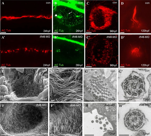
Ciliary defects in zebrafish ift46 morphants. Immunofluorescence with antibody against acetylated α-tubulin showing cilia in the pronephric ducts, (scale bar, 10 µm) (A–A′), otic vesicle, (scale bar, 10 µm) (B–B′), olfactory placode, (scale bar, 10 µm) (C–C′) and a lateral line hair cell, (scale bar, 5 µm) (D–D′). The ift46 morphants exhibit shortened or fewer cilia compared with the control fish in all of these organs. Arrow head: tether cilia. Arrow: short cilia. (E–F′) SEM images showing the cilia in the olfactory placode of control fish (E, E′) and ift46 morphants (F, F′) at 5 dpf. Scale bar, 40 µm (E) and (F), 4 µm (E′) and (F′) (G–H′) TEM images of cilia in zebrafish pronephric ducts at 72 hpf. Ultrastructure of normal pronephric cilia shows 9+2 microtubule architecture at 72 hpf which is intact in ift46 morphants. (G′, H′) Representative views of individual cilium at higher magnification. Scale bar, 5 µm (G) and (H), 100 µm (G′) and (H′). |

Overexpression of IFT46 induces apoptosis in zebrafish embryos. (A, B) Control and human IFT46 mRNA (100 pg/embryos) injected embryo stained with acridine orange at 24 hpf. overexpression of IFT46 leads to increase of apoptosis in developing embryo. (C) Temporal expression profile of ift46 by RT-PCR. The ift46 transcript is detected from 4-cell stage to 72 hpf in zebrafish. Both maternal and zygotic expression of ift46 is detected. β-actin is a loading control. (D and E) ift46-GFP expression is completely inhibited when co-injection with ift46 translation blocking morpholino in zebrafish embryos, showing the efficacy of the ift46-MO. EXPRESSION / LABELING:
|

The ift46 intraflagellar transport complex B protein 46 C-terminal domain is sufficient for ift46 function. (A) Diagram of ift46 and deletion mutant structures. Numbers indicate the amino acid positions in ift46 protein. (B) Quantification of the rescue of ift46 morpholino phenotypes by co-injection of human and zebrafish mRNA. Control (n=144), ift46-MO (n=180), IFT46 mRNA+ift46-MO (n=120), zebrafish ift46 mRNA+ift46-MO (n=108). Statistical significance of pairwise comparisons are shown (***P<0.001; Studentós t-test). (C-H) Representative images of zebrafish larvae following co- injection of ift46-MO and ift46 encoding mRNAs. |

The organ laterality defect is not disrupted in zebrafish ift46 morphants. (A–C′) The ift46 morphant embryos have no left–right patterning defect visualized by whole-mount in situ hybridization for lft1 and lft2 (D–E′). The ift46 morphants does not have any abnormal heart looping and gut laterality with whole-mount in situ hybridization for cmlc and foxA3. (A, artery; I, intestine; L, liver; P, pancreas; V, vein). (F–F′) Expression of charon in Kupfferós vesicle in the control and the ift46 morphants are comparable. (G–G′) Visualization of cilia with immunostaining of acetylated α-tubulin in the Kupfferós vesicle in both control and the ift46 morphants at 6-somite stage. Scale bar, 20 µm. (H and I) Organ asymmetry scored by cmlc2 (Control (n=56) and ift46 morphants (n=85)) and foxA3 (Control (n=68) and ift46 morphants (n=81)) expression. (J and K) Quantification of the number and length of the KV cilia shows no statistical differences between the control and the ift46 morphant embryos. PHENOTYPE:
|
Reprinted from Developmental Biology, 400(2), Lee, M.S., Hwang, K.S., Oh, H.W., Ji-Ae, K., Kim, H.T., Cho, H.S., Lee, J.J., Yeong Ko, J., Choi, J.H., Jeong, Y.M., You, K.H., Kim, J., Park, D.S., Nam, K.H., Aizawa, S., Kiyonari, H., Shioi, G., Park, J.H., Zhou, W., Kim, N.S., Kim, C.H., IFT46 plays an essential role in cilia development, 248-57, Copyright (2015) with permission from Elsevier. Full text @ Dev. Biol.