
- Title
-
Molecular Description of Eye Defects in the Zebrafish Pax6b Mutant, sunrise, Reveals a Pax6b-Dependent Genetic Network in the Developing Anterior Chamber
- Authors
- Takamiya, M., Weger, B.D., Schindler, S., Beil, T., Yang, L., Armant, O., Ferg, M., Schlunck, G., Reinhard, T., Dickmeis, T., Rastegar, S., Strähle, U.
- Source
- Full text @ PLoS One
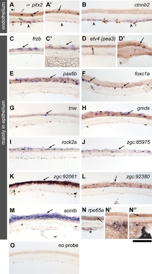
In situ mRNA expression analysis in the adult zebrafish cornea. Two major classes of expression patterns are shown for selected genes: genes mainly expressed in the corneal endothelium (paired-like homeodomain 2 (A-A′) and catenin, beta 2 (B)) and mainly in the corneal epithelium (frizzled-related protein (C), ets variant 4 (previously known as pea3, D-D′), paired box 6b (E), forkhead box C1a (F), tenascin W (G), GDP-mannose 4,6-dehydratase (H), rho-associated, coiled-coil containing protein kinase 2a (I), zgc:85975 (J), zgc:92061 (K), zgc:92380 (L), scinderin like b (M) and retinal pigment epithelium-specific protein 65a (N-N′′). A control without probe is shown in the panel O. Filled arrows, open arrows, arrowheads and asterisks indicate expression in the corneal epithelial, stromal, endothelial layers and the annular ligament, respectively. The black “flakes” (indicated by p in the panels of A, B, K and N′′) are derivatives of pigmentation unavoidably introduced during cryosectioning and its post-processing. Scale bar: 50 µm. EXPRESSION / LABELING:
|

Genes expressed in the cornea are also expressed in the retina of the adult eye. (A) Analysis of genes and their expression in structures of the adult zebrafish eye. The 32 genes examined are listed along the vertical axis. The examined anatomical structures are shown in the schematic and listed on the horizontal axis. Anatomical structures examined are inner nuclear layer of the retina (INL), iris stroma (irsStr), outer nuclear layer and outer plexiform layer (ONL-PR), ganglion cell layer (GCL), ciliary marginal zone of the retina (CMZ), corneal epithelium (crnEpi), annular ligament (AL), sclera (scl), corneal stroma (crnStr), peripheral region of the corneal endothelium (crnEndperi) and central region of the corneal endothelium (crnEndcen). (B-E): Examples of genes expressed in the cornea and the retina. in situ gene expression patterns of foxc1a (B, B′), pax6b (C, C′) and pitx2 (D, D′) are shown in the adult cornea (B-D) and retina (B′-D′), with tissue processed without antisense mRNA probe as a negative control (E, E′). Genes expressed in either the corneal epithelium (arrows) or the corneal endothelium (asterisk) are also expressed in the retina. rpe: retinal pigment epithelium; on: outer nuclear layer; in: inner nuclear layer; gcl: ganglion cell layer. Scale bar: 50 µm. EXPRESSION / LABELING:
|

Comparison of gene expression in the cornea of wild type zebrafish at 7 dpf (A-O) and 1-month stage (A′-O′). Transverse sections with dorsal up. (A) Negative controls without probe showed no staining at both stages, except for background staining in the lens (ln, outlined by a stippled line). p: pigmentation in the iris and the retinal pigment epithelium. (B-K′) Examples of genes expressed at both 7 dpf and 1-month stage are shown. A subset of genes (B-F′) is mainly expressed in the corneal epithelium (arrowheads) and another subset (G-K′) shows expression in the corneal endothelium (arrows). (L-O′) Examples of genes that are mainly expressed at the 1-month stage. A subset of genes (M-O′) is expressed in the corneal epithelium (arrowheads) and another subset (L-L′) in the corneal endothelium (arrows). The gene symbols are indicated in the bar above each pair of sections. Scale bar: (A-O) 80 µm; (A′-O′) 100 µm. EXPRESSION / LABELING:
|

pax6b mutants (also named sunrise) present an abnormal anterior chamber with severe corneal endothelium defects. (A-D) pax6b in situ expression at 28 hpf (A), 3 dpf (B), 7 dpf (C) and 30 dpf (D). Transverse sections through the eye are shown with the ventral side left. (A) At 28 hpf, only a marginal level of pax6b transcripts is found in the ocular mesenchyme (arrow), well in contrast to high expression levels in the developing retina (Re) and the lens (Ln, stippled circle). (B-D) At 3 dpf, cells in the developing anterior chamber start to express pax6b (arrows in B), and its expression is maintained at 7 dpf (arrow in C) and 30 dpf (arrow in D). At 30 dpf, the corneal epithelium expresses pax6b transcripts (arrowhead, D) and its expression is maintained in the adult (Fig. 2E). (E-H) The anterior chamber phenotype of a pax6b mutant. (E-F) At 7 dpf, wildtype embryos have a well-formed anterior chamber (asterisk, E), while pax6b mutants show almost no anterior chamber (asterisk, F) and severely malformed corneal structures (black arrow, F) appear to be directly in contact with the lens. (G-H) Ultrastructure analysis of the cornea at 7 dpf. Wildtype embryos show the corneal endothelium (En, pseudo-coloured in blue) as consistent monolayer of cells. pax6b mutants lack a well formed corneal endothelium, and abnormal mesenchymal cells (pseudo-coloured in blue) with cellular protrusions (white arrow) were observed instead. Vacuolar deposits were present in the lens (asterisk). Ep: corneal epithelium, St: corneal stroma. Ln: lens. Scale bars: (A-D) 35 µm, (E-F) 93 µm and (G-H) 1.0 µm. EXPRESSION / LABELING:
PHENOTYPE:
|
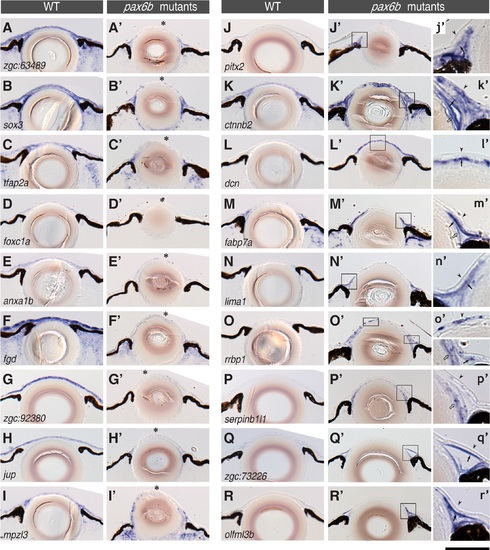
Gene expression in the cornea is severely affected in pax6b mutants. The distribution of the mRNA of cornea genes was compared at 7 dpf between wildtype (A-R) and pax6b mutant embryos (A′-R′). Each panel represents a transverse section through the eye and the lens. The gene transcripts analysed by in situ hybridization are zgc:63489 (A-A′), sox3 (B-B′), tfap2a (C-C′), foxc1a (D-D′), anxa1b (E-E′), fgd (F-F′), zgc:92380 (G-G′), jup (H-H′), mpzl3 (I-I′), pitx2 (J-J′), ctnnb2 (K-K′), dcn (L-L′), fabp7a (M-M′), lima1 (N-N′), rrbp1 (O-O′), serpinb1l1 (P-P′), zgc:73226 (Q-Q′) and olfml3b (R-R′). (A-I′) Examples for the loss of gene expression in the pax6b mutant (asterisks). (J-R′) Genes whose expression is ectopically induced in the pax6b mutant. The insets (j′-r′) represent magnified views of corresponding cornea regions (rectangular areas in J′-R′); arrowheads, filled arrows and open arrows indicate the corneal epithelium, the endothelium and the lens epithelium. The ectopic expression in pax6b mutants was mostly observed in the endothelial layer or in the iridocorneal angle. In contrast, the loss of gene expression in the pax6b mutant was mostly detected in the epithelial layer (asterisks in A-I). Scale bar: 64 µm (A-R′) and 20 µm (j′-r′). EXPRESSION / LABELING:
|