
- Title
-
The secreted factor Ag1 missing in higher vertebrates regulates fins regeneration in Danio rerio
- Authors
- Ivanova, A.S., Shandarin, I.N., Ermakova, G.V., Minin, A.A., Tereshina, M.B., Zaraisky, A.G.
- Source
- Full text @ Sci. Rep.
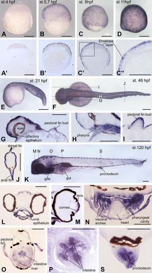
Spatial expression pattern of DAg1 in Danio rerio embryos as revealed by in situ hybridization. (A–D and E). Up to 21hpf, DAg1 is expressed only in the superficial enveloping layer. (A′–C′). Histological sections of embryos shown on A–C. (C′′). Enlarged fragment framed on C′ by dotted line. (F–J). Dorsal view of the embryo at hatching stage (F) and histological sections (G–J) of sibling embryos corresponding to the planes indicated by dotted lines on F. (K–S). Left side view of embryo at 5 days stage (K) and histological sections (L–S)of sibling embryos corresponding to the planes indicated by dotted lines on K. Scale bar everywhere is 200µm. EXPRESSION / LABELING:
|
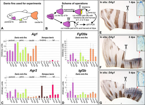
Analysis of Ag1, Agr2, Fgf20a and IGF2b expression in the regenerating fins. (A and B). Schemes of experiments. Tissue pieces of hindlimbs and tails of stage 52 Xenopus laevis tadpoles were collected as was previously described5. Drawings for these figures were done by MBT and AGZ. (C and D). QRT-PCR analysis of Ag1 and Agr2 (C) and Fgf20a and Igf2b (D) expression in the intact and regenerating of fins of adult Danio rerio and in hindlimbs and tails of stage 52 Xenopus laevis tadpoles. All graphs represent means of triplicate experiments. Bars indicate standard deviations. The geometric mean of expression of Danio rerio and Xenopus laevis ornithine decarboxylase (ODC) and elongation factor 1alpa (EF-1alpha) was used for normalization of experimental values (see Materials and Methods for details). The value of normalized PCR signal in "m" sample taken from pectoral fin was taken as an arbitrary unit in each series. (E–G). In situ hybridization of regenerating caudal fins with the probe to DAg1 at 1, 2 and 5dpa. Photos on insets demonstrate histological sections of regenerating fins at levels whose approximate positions are indicated by red dotted arrowed lines. Black dashed lines indicate places of amputations. Abbreviations: bl - blastema, ep - epidermis. EXPRESSION / LABELING:
|
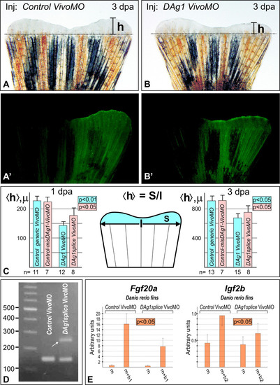
Blocking of DAg1 mRNA translation by Vivo-morpholino injections results in retardation of the caudal fin regeneration. (A and A′). Caudal fin injected by the control-generic Vivo-morpholino mixed with FLD in the right side. No retardation of regeneration is seen on the injected side. Dashed line indicates the level of amputation. Scale bar 1mm. (B and B′). Caudal fin injected by the DAg1 Vivo-morpholino mixed with FLD in the right side. Note retardation of regeneration of the injected side. Scale bar 1mm. (C). Quantification of the mean height of the regenerating part of caudal fins injected by control-generic Vivo MO or DAg1 Vivo MO at 1dpa (left) and 3dpa (right). Numbers of the injected fins are indicated by n. The schema in the middle demonstrates how mean height <h> was calculated. Drawing on this figure was done by AGZ. (D). RT-PCR analysis of DAg1 transcripts in 1dpa regenerating caudal fins injected with either control-generic Vivo MO or DAg1splice Vivo MO. White arrow indicates additional band generated by DAg1 un-spliced transcript. (E). QRT-PCR analysis of Fgf20a and Igf2b expression in the regenerating caudal fin at 1dpa and 2dpa respectively (at these days the expression levels of Fgf20a and Igf2b reach maximal values, see Fig. 3D) maximal expression levels of these genes injected with either control-generic Vivo MO or DAg1splice Vivo MO. For scheme of experiments and abbreviations see picture D and legends on Fig. 3). |
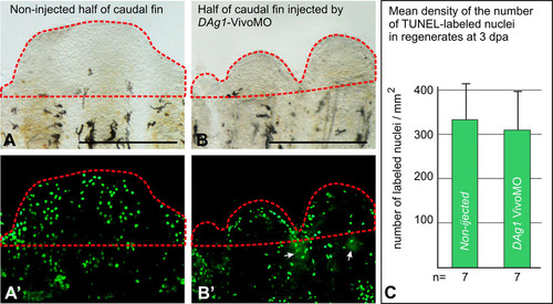
TUNEL assay of apoptosis in the regenerating caudal fins injected with Vivo MO. (A and A′). Revealing by TUNEL assay of apoptotic nuclei in regenerating part (framed by red dashed line) of the non-injected side of the caudal fin at 3dpa. Scale bar 500µm. (B and B′). Revealing by TUNEL assay of apoptotic nuclei in regenerating part (framed by red dashed line) of the side of caudal fin injected by DAg1Vivo MO at 3dpa. White arrows indicate traces of FLD co-injected with MO. Scale bar 500µm. (C). Quantification of TUNEL-labeled nuclei in the regenerating parts (regenerates) of caudal fins non-injected and injected by DAg1 Vivo MO. |

Control in situ hybridization. A. In situ with antisense DAg1 probe on the 11 hpf embryo cut sagittally before hybridization. DAg1 expression is seen only in the superficial layer of cells, but not in cells of deeper layers. This confirms that the in situ hybridization signal, which was observed primarily in the superficial cells of embryos hybridized in whole-mount, was not the result stipulated by a poor penetration of DAg1 probe into the depth of embryo. Anterior to the left, dorsal side up. B and C. No signal was detected when embryos at 8 and 36 hpf were hybridized in whole mount with sense probe to DAg1. This result confirms specificity of the signal observed with antisense DAg1 probe. On C animal pole to the top. On C anterior to the left, dorsal side up. Scale bar everywhere is 200 µm |
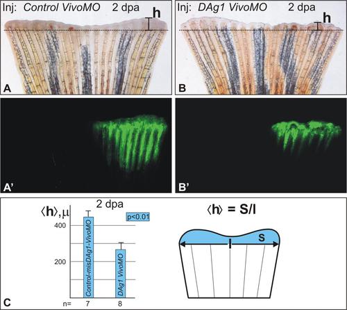
Comparison of effects of DAg1 and control mismatch DAg1MO. A and A′. Caudal fin injected by the control-misDAg1 Vivo-MO mixed with FLD in the right side. No retardation of regeneration is seen on the injected side. Dashed line indicates the level of amputation. B and B′. Caudal fin injected by the DAg1 Vivo-morpholino mixed with FLD in the right side. Note retardation of regeneration of the injected side. C. Quantification of the mean height of the regenerating part of caudal fins injected by controlmisDAg1 Vivo-MO or DAg1 Vivo MO at 2 dpa. Numbers of the injected fins are indicated by n. The schema in the middle demonstrates how mean height |