
- Title
-
Fgf16 Is Required for Specification of GABAergic Neurons and Oligodendrocytes in the Zebrafish Forebrain
- Authors
- Miyake, A., Chitose, T., Kamei, E., Murakami, A., Nakayama, Y., Konishi, M., Itoh, N.
- Source
- Full text @ PLoS One
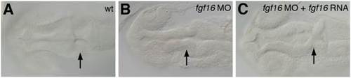
Morphology of the brain in fgf16 morphants. Dorsal views of wild-type (A), fgf16 MO-injected (B), and fgf16 MO- and fgf16 RNA-injected (C) embryos at 24 hpf. Arrows indicate the MHB constriction. PHENOTYPE:
|
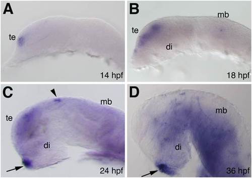
Expression pattern of fgf16 in the brain during zebrafish embryonic development. (A–D) Expression pattern of fgf16 in zebrafish embryos at the indicated stages as detected by whole-mount in situ hybridization. Lateral views with anterior to the left and dorsal to the top. Arrows and arrowheads indicate the pituitary gland and epiphysis, respectively. di, diencephalon; mb, midbrain; te, telencephalon. EXPRESSION / LABELING:
|

Comparison of cell proliferation and cell death patterns in control embryos and fgf16 morphants. (A, B) Control embryos (A) and embryos injected with fgf16 MO (B) were stained using an anti-H3P antibody. Panels show representative horizontal sections of the head region at 24 hpf. (C, D) The percentage of proliferating cells labelled with the anti-pH3 antibody in the forebrain (C) and midbrain (D) of control embryos and embryos injected with fgf16 MO. Results are the mean ± S.D. for three independent sections from three embryos. The significance of differences in mean values was assessed with the Student’s t-test. Asterisks indicate significant differences from the control (*P<0.05). The forebrain (fb) and midbrain (mb) regions, which we defined in the sections, are separated by black lines. Scale bar: 25 µm. PHENOTYPE:
|
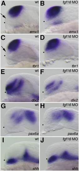
Telencephalic and diencephalic gene expression in the fgf16 morphants. The expression of emx1 (A, B), tbr1 (C, D), dlx2 (E, F), pax6a (G, H), and shh (I, J) in wild-type embryos (A, C, E, G, I) and fgf16 morphants (B, D, F, H, J) at 24 hpf. Arrows in panels A and C indicate the subpallial telencephalon, which was negative for emx1 or tbr1. Dots indicate the boundary between the telencephalon and ventral diencephalon. Lateral views with anterior to the left and dorsal to the top. EXPRESSION / LABELING:
|
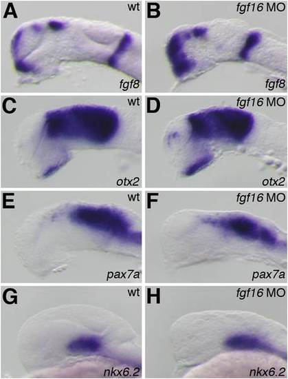
Gene expression in the midbrain and MHB of the fgf16 morphants. The expression of fgf8 (A, B), otx2 (C, D), pax7a (E, F), and nkx6.2 (G, H) in wild-type embryos (A, C, E, G) and fgf16 morphants (B, D, F, H) at 24 hpf. Lateral views with anterior to the left and dorsal to the top. EXPRESSION / LABELING:
|

Effects of fgf16 on the development of glutamatergic neurons, GABAergic interneurons, and oligodendrocyte progenitor cells. (A–D) The expression of ngn1 (A, B) and isl1 (C, D) in wild-type embryos (A, C) and fgf16 morphants (B, D) at 24 hpf. Lateral views with anterior to the left and dorsal to the top. (E–J) The expression of gad1 (E, F), olig2 (G, H), and slc17a6a (I, J) in wild-type embryos (E, G, I) and fgf16 morphants (F, H, J) at 28 hpf. Lateral views with anterior to the left and dorsal to the top. |

Effects of fgf16 on the differentiation of GABAergic interneurons and oligodendrocytes. (A, B) Dorsal views of wild-type embryos (A) and fgf16 morphants (B), labeled to show GABA immunoreactivity at 3 dpf. (C, D) The expression of plp in wild-type embryos (C) and fgf16 morphants (D) at 4.5 dpf. Lateral views with anterior to the left and dorsal to the top. EXPRESSION / LABELING:
PHENOTYPE:
|

Interactions between fgf16 and Hh signaling in the forebrain and midbrain. The expression of fgf16 at 16 (A, B) and 24 (C, D) hpf in wild-type embryos treated with 0.95% ethanol (A, C) or cyclopamine (B, D). Arrows in panels A and C indicate fgf16 expression in the telencephalon. The arrowhead in panel C indicates fgf16 expression in the midbrain. Lateral views with anterior to the left and dorsal to the top. EXPRESSION / LABELING:
|
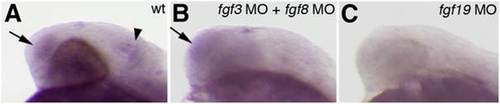
Interactions between fgf3, fgf8, fgf19 and fgf16. The expression of fgf16 at 24 hpf in wild-type embryos (A) and embryos injected with fgf3 MO and fgf8 MO (B), and fgf19 MO (C). Arrows in panels A and B indicate fgf16 expression in the telencephalon. The arrowhead in panel A indicates fgf16 expression in the midbrain. Lateral views with anterior to the left and dorsal to the top. |

Apoptosis in the brain of fgf16 morphants. At 24 hpf, apoptotic cells in the brain of the wild-type (A) and fgf16 MO1-injected (B) embryos were marked via TUNEL. Lateral views with anterior to the left and dorsal to the top. PHENOTYPE:
|

Oligodendrocyte differentiation in the hindbrain of fgf16 morphants. (A, B) Dorsal views of wild-type embryos (A) and fgf16 morphants (B), labeled to show CC1/APC immunoreactivity at 4.5 dpf. |