
- Title
-
Dynamic miRNA Expression Patterns During Retina Regeneration in Zebrafish: Reduced Dicer or miRNA Expression Suppresses Proliferation of Müller Glia-Derived Neuronal Progenitor Cells
- Authors
- Rajaram, K., Harding, R.L., Bailey, T., Patton, J.G., Hyde, D.R.
- Source
- Full text @ Dev. Dyn.
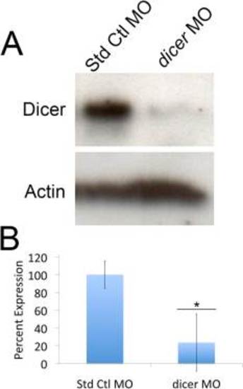
Morpholino-mediated knockdown of Dicer protein expression. Lissamine-tagged Standard Control morpholino (Std Ctl MO) or dicer morpholino were intravitreally injected and electroporated into dark-adapted albino retinas. After 35h constant light, dosal retinas were isolated and the expression of Dicer and Actin were analyzed by immunoblots (A). After incubation with the secondary antibody, the membrane was separated to allow a longer exposure of the Dicer region relative to the Actin region. After adjusting for the difference in the amount of the Actin protein in the two samples, the dicer morphant retinas possessed 77% less Dicer protein relative to the Standard Control morphant retina (B, *P<0.05, n=5). |
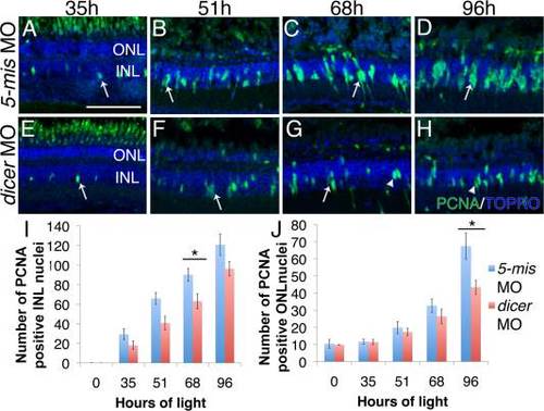
Dicer knockdown decreased INL proliferation in the light-damaged retina. Lissamine-tagged dicer 5-base mismatch (5-mis) control morpholino or dicer morpholino were intravitreally injected and electroporated into dark-adapted adult albino zebrafish. Retinas were collected at 0, 35, 51, 68, and 96h of light damage and immunostained with anti-PCNA (green) antibodies and TOPRO3 nuclear stain (blue). A, E: At 35h, single PCNA-positive Müller glia were observed in the INL of dicer 5-mis control and dicer morphant retinas (arrows). B, F: Both dicer 5-mis control and dicer morphant retinas contain doublet nuclei at 51h of light damage (arrows). C: Clusters of proliferating progenitor cells are present in the INL of dicer 5-mis control retinas (arrow) at 68h of light damage. G: Single (arrow) or doublet (arrowhead) nuclei predominated in dicer morphant retinas. D: Columns of proliferating progenitors were observed in the INL of dicer 5-mis control morphant retinas at 96h of light damage. H: At 96h of light damage, doublet nuclei (arrowhead) predominated the INL of dicer morphants. I: Significantly fewer PCNA-positive INL cells were present in dicer morphant retinas compared to dicer 5-mis control morphant retinas beginning at 68h of light damage. J: dicer morphant retinas contained significantly fewer PCNA-positive ONL cells at 96h of light damage. dicer 5-mis MO, dicer 5-base mismatch control morphant; dicer MO, dicer morphant; INL, inner nuclear layer; ONL, outer nuclear layer. Scale bar in A = 50 µm and applies to B–H; data represent mean ± s.e.m; *P< 0.05 using two-way ANOVA with a Tukey′s post-hoc test, n=11. |
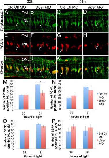
Dicer knockdown does not affect Müller glia proliferation in light-damaged retinas. Adult albino Tg(gfap:egfp)nt11 zebrafish were intravitreally injected and electroporated with standard control or dicer morpholino prior to the start of light damage. A–L: EGFP-positive Müller glia (arrows) were observed in comparable numbers between dicer and standard control morphants at all time points (A–D, I–L, O, P). E,F: PCNA-positive INL cells were observed as mainly single nuclei in dicer and standard control morphant retinas at 35h of light damage (arrows). IJ: Overlay of these images revealed that many Müller glia co-labeled with PCNA as they re-entered the cell cycle. G: Standard control morphant retinas at 51h of light damage exhibited doublets or early columns of PCNA-positive INL cells. H: Single PCNA-positive INL cells rather than doublets predominated in dicer morphant retinas (arrows). M–N: Differences in proliferation in dicer morphants resulted in significantly fewer PCNA-positive INL cells at 51h of light, but no difference in PCNA-positive ONL cells. Std Ctl MO, Standard Control morphant; dicer MO, dicer morphant; INL, inner nuclear layer; ONL, outer nuclear layer. Scale bar in A = 50 µm and applies to B–L; *P<0.05 using two-way ANOVA with a Tukey′s post-hoc test, n=7. |
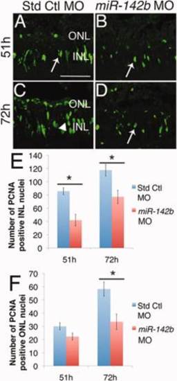
Knockdown of miR-142b reduces proliferation. Adult albino zebrafish were intravitreally injected and electroporated with either standard control or miR-142b morpholino prior to the start of light damage. A,B: After 51h of constant light, doublet PCNA-positive INL cells (arrows) were present in both the standard control and miR-142b morphant retinas. C,D: After 72h, PCNA-positive INL cell clusters are present in standard control morphants (C, arrowhead), but only doublet or fewer PCNA-positive INL cells (D, arrow) remained in miR-142b morphant retinas. E,F: miR-142b knockdown significantly reduced the number of PCNA-positive INL cells at both 51 and 72h and ONL cells at 72h. INL, inner nuclear layer; ONL, outer nuclear layer. Scale bar in A = 50 µm and applies to B–D; data represent mean ± s.e.m. *P< 0.05 using two-way ANOVA with Tukey′s post-hoc test, n=6. |
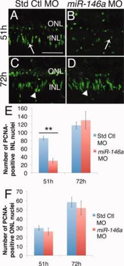
Knockdown of miR-146a reduces proliferation at 51h. Adult albino zebrafish were intravitreally injected and electroporated with either standard control or miR-146a morpholino prior to the start of light damage. A,B: After 51h of constant light, doublet PCNA-positive INL cells are present in standard control morphants (arrow), but mainly single PCNA-positive INL cells are present in miR-146a morphants (arrow). C,D: By 72h, both standard control and miR-146a morphants contained columns of INL proliferating nuclei (arrowheads). E,F: miR-146a knockdown significantly reduced the number of proliferating nuclei at 51h, but not at 72h. There was not a significant difference in the number of PCNA-positive INL cells at either 51h or 72h between the miR-146a and standard control morphant retinas. INL, inner nuclear layer; ONL, outer nuclear layer. Scale bar in = 50 µm and applies to B–D. Data represent mean ± s.e.m. **P< 0.01 using two-way ANOVA with Tukey′s post-hoc test, n=6. |
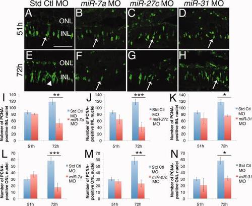
Knockdown of miR-7a, miR-27c and miR-31 reduced proliferation at 72h. Adult albino zebrafish were intravitreally injected and electroporated with either standard control miR-7a, miR-27c or miR-31 morpholino prior to the start of light damage. A–D: After 51h of light, standard control, miR-7a, miR-27c, and miR-31 morphant retinas contained PCNA-positive nuclei in the INL (arrows). E: At 72h, standard control morphant retinas contain columns of INL proliferating nuclei (arrowhead). F–H: At 72h, miR-7a, miR-27c and miR-31 morphant retinas contained mainly doublet INL and single nuclei (arrows). I–K: There were no significant differences in the number of PCNA-positive INL cells at 51h between the standard control morphant retina and the miR-7a (I), miR-27c (J), and miR-31 (K) morphant retinas. In contrast, significantly fewer PCNA-positive INL cells were present in all three miRNA morphant retinas relative to standard control morphant retinas at 72h. L–N: There were no significant differences in the number of PCNA-positive ONL cells at 51h between the standard control morphant retina and the miR-7a (L), miR-27c (M), and miR-31 (N) morphant retinas. In contrast, significantly fewer PCNA-positive INL cells were present in all three miRNA morphant retinas compared to standard control morphant retinas at 72h. INL, inner nuclear layer; ONL, outer nuclear layer. Scale bar in A = 50 µm and applies to B–H. Data represent mean ± s.e.m. *P< 0.05; ** P < 0.01; *** P <0.001 using two-way ANOVA with Tukey′s post-hoc test, n=6. |
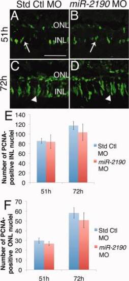
Knockdown of miR-2190 does not affect proliferation. Adult albino zebrafish were intravitreally injected and electroporated with either standard control or miR-2190 morpholino prior to the start of light damage. A,B: After 51h of constant light, standard control and miR-2190 morphant retinas both display PCNA-positive doublet INL proliferating nuclei (arrows). C,D: After 72h of light, standard control and miR-2190 morphant retinas display clusters of INL nuclei (arrowheads). E,F: Statistically equivalent numbers of INL and ONL PCNA-positive nuclei are present in standard control and miR-2190 morphant retinas. INL, inner nuclear layer; ONL, outer nuclear layer. Scale bar in A = 50 µm and applies to B–D; data represent mean ± s.e.m. P > 0.05 using two-way ANOVA with Tukey′s post-hoc test, n=6. EXPRESSION / LABELING:
|