
- Title
-
Tip-link protein protocadherin 15 interacts with transmembrane channel-like proteins TMC1 and TMC2
- Authors
- Maeda, R., Kindt, K.S., Mo, W., Morgan, C.P., Erickson, T., Zhao, H., Clemens-Grisham, R., Barr-Gillespie, P.G., Nicolson, T.
- Source
- Full text @ Proc. Natl. Acad. Sci. USA
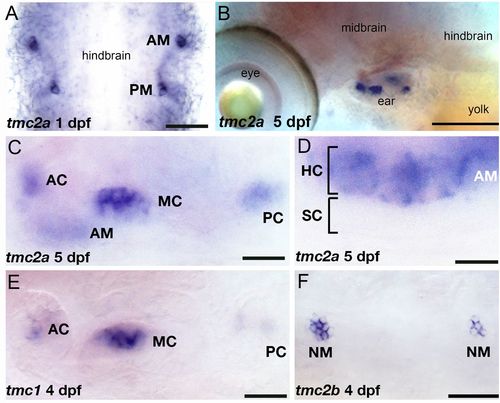
Embryonic and larval expression pattern of the tmc1/2 genes. In situ hybridization with specific probes for each transcript was performed on stages 1–5 dpf. Expression of tmc2a at 1 dpf (A, flat mount) and 5 dpf (B). Higher magnification view of tmc2a expression in the inner ear (C) and anterior macula at 5 dpf (D). Label is restricted to the upper hair cell layer of the neuroepithelium. (E) Expression of tmc1 at 4 dpf (focal plane: cristae). (F) Expression of tmc2b in neuromasts, 4 dpf. AC, anterior crista; AM, anterior macula; HC, hair cells; MC, medial crista; NM, neuromast; PC, posterior crista; SC, supporting cells. [Scale bars: 50 μm (A); 130 μm (B); 22 μm (C and E); 12 μm (D); 43 μm (F).] |
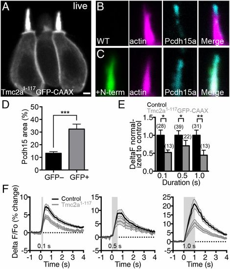
Expression of the N-terminal fragment of Tmc2a decreases mechanically evoked responses in zebrafish hair cells. (A) Live confocal image (0.35-μm z sections) of mosaic expression of Tmc2a1–117-GFP-CAAX in ampullary hair cells at 3 dpf. Two mature hair cells are shown in the focal plane. (B–D) Altered distribution of Pcdh15a in hair cells expressing Tmc2a1–117-GFP-CAAX (4 dpf). Control hair bundle (B) and Tmc2a1–117-GFP-CAAX–positive hair bundle (C) were labeled with anti-GFP antibody (green), phalloidin (magenta), and anti-Pcdh15a antibody (cyan). (D) Quantification of the increase in pixel area of Pcdh15a immunolabel within hair bundles (n = 19 hair bundles for each condition; Student t test). [Scale bar: 1.75 μm (A); 1 μm (B and C)]. (E) Response magnitude in Tmc2a1–117-GFP-CAAX–expressing cells normalized to control signals. The number of cells is indicated above the bars. A nonparametric Mann–Whitney U test (unpaired) was used to compare differences between Tmc2a1–117-GFP-CAAX and control hair cell calcium responses. (F) Averaged traces of mechanically evoked calcium responses are plotted as a percentage (ΔF F0/F0 × 100) and as a function of time in response to 0.1-, 0.5-, and 1.0-s steps. Duration of the step stimulus is indicated by the gray rectangles. The gray shading around traces in F and error bars in E indicates SEM. EXPRESSION / LABELING:
PHENOTYPE:
|
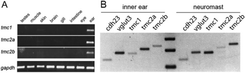
Expression of tmc1, tmc2a, and tmc2b transcripts in adult and larval tissues. (A) RT-PCR revealed products for all three genes in the inner ear. Lanes: 1, testes; 2, muscle; 3, skin; 4, brain; 5, gills; 6, intestine; 7, eye; 8, ear. cDNA for gapdh was used as a positive control. (B) RT-PCR of cDNA isolated from larval inner ears and lateral-line neuromasts. Control reactions include amplification of hair cell-specific cdh23 and vglut3 cDNA. |

N-terminal fragments of distantly related Tmcs accumulate within the cell body of hair cells. (A–C) Live images of ampullary hair cells. (A) Tmc4 (1–141 aa) and (B) Tmc5 (1–130 aa) were tagged with GFP-CAAX and mosaically expressed in hair cells using the myo6b promoter. In both low and high expressing hair cells, GFP signal is associated either with intracellular membrane compartments or the basolateral membrane and mostly excluded from hair bundles. Some faint bundle label is seen in the cell of A (n e 24 hair cells for each fragment). (C) Example of morphological defects in a hair cell expressing high levels of Tmc2a1–117-GFP-CAAX. The hair bundle appears amorphous (arrow), and blebbing of the plasma membrane (arrowhead) and unusually long apical processes are evident. In contrast to Tmc4 and Tmc5, large brightly labeled internal compartments were not observed. Cells with morphological defects were excluded from our immunohistochemical and physiological experiments. (Scale bar: 5 μm.) |

Immunolabeling of Pcdh15a in wild-type siblings and pcdh15apsi7 mutants. (A) Sequence of the psi7 allele, which encodes splice site mutation causing a frameshift following exon 7. (B) The predicted product is a short N-terminal fragment of Pcdh15a. (C) Immunolabeling with a monoclonal antibody against Pcdh15a. Actin filaments were labeled with rhodamine–phalloidin. (Right) Note the splaying of stereocilia, which is typical for mutants expressing strong alleles of pcdh15a (1). (Scale bar: 5 μm.) |
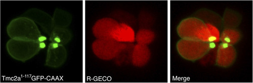
Example of the Red-GECO1 indicator in lateral-line hair cells. Top-down view of the mosaic expression of Tmc2a1–117-GFP-CAAX in hair cells expressing R-GECO1 at 3 dpf. In this example, four out of eight cells are GFP-CAAX positive. Note the four bright bundles near the center of the rosette. Most of the GFP signal is concentrated at the plasma membrane. Mature GFP-negative cells served as controls, allowing direct comparison of cells within the same neuromast. |
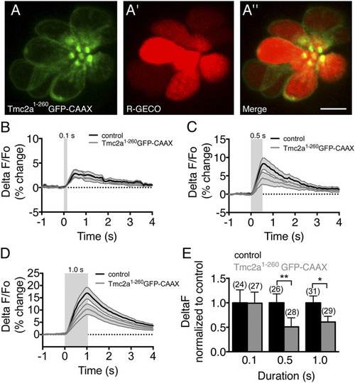
Decreased mechanosensitive responses of hair cells stably expressing Tmc2a1–260-GFP-CAAX at 3 dpf. (A–A′′) Example of expression of the 260-aa N-terminal fragment in lateral-line hair cells. The bright oval-shaped puncta of exogenous Tmc2a fragments are enriched in apical hair bundles at the center of the rosette, although more label is present in the soma in comparison with the 117-aa fragment (Fig. S9). (Scale bar: 5 μm.) (B–D) Calcium transients in lateralline hair cells in response to 0.1-, 0.5-, and 1.0-s deflections. (E) Summary of responses normalized to transgenic R-GECO1 control hair cells. EXPRESSION / LABELING:
PHENOTYPE:
|