
- Title
-
Regenerative responses after mild heart injuries for cardiomyocyte proliferation in zebrafish
- Authors
- Itou, J., Akiyama, R., Pehoski, S., Yu, X., Kawakami, H., Kawakami, Y.
- Source
- Full text @ Dev. Dyn.

Comparison of injury to the myocardial layer by different injury methods. A–D: In situ hybridization of cmlc2a (purple) after various injuries to the heart. E–S: Immunostaining of MHC (brown) coupled with fibrin detection (red) after various injuries to the heart. Sham operation (A, E), scratch-induced injury (B, F, I, L, O), puncture-induced injury (C, G, J, M, P, R), and resection-induced injury (D, H, K, N, Q, S) were performed. Arrowheads point to fibrin clot. Time points after injury are indicated. Brackets indicate the injury sites. Scale bar = 50 µm. |
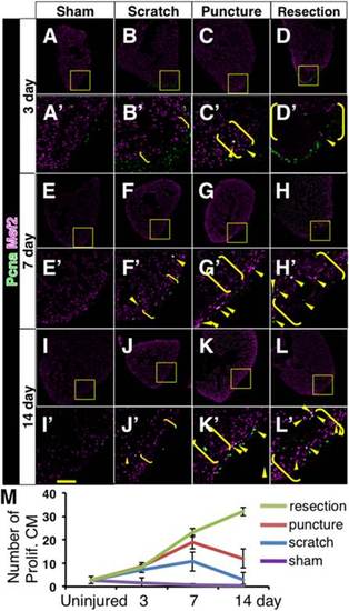
Cardiomyocyte proliferation after different damages to the heart. A–L, A′–L′: Representative images of MEF2 (magenta) and PCNA (green) immunostaining at 3 dpi (A–D, A′–D′), 7 dpi (E–H, E′–H′), and 14 dpi (I–L, I′–L′) after sham operation (A, A′, E, E′, I, I′), scratch- (B, B′, F, F′, J, J′), puncture- (C, C′,G, G′, K, K′), and resection- (D, D′, H, H′, L, L′) induced injury. Yellow arrowheads point to proliferating cardiomyocytes, observed as white signals. A′–L′ show close-up images of the boxed areas in A–L. Brackets indicate the injury sites. Scale bar = 50 µm. M: A graphic representation of the number of proliferating cardiomyocytes at indicated time points after different injury methods. Shown are average ± S.D. (n=3). CM, cardiomyocyte. |
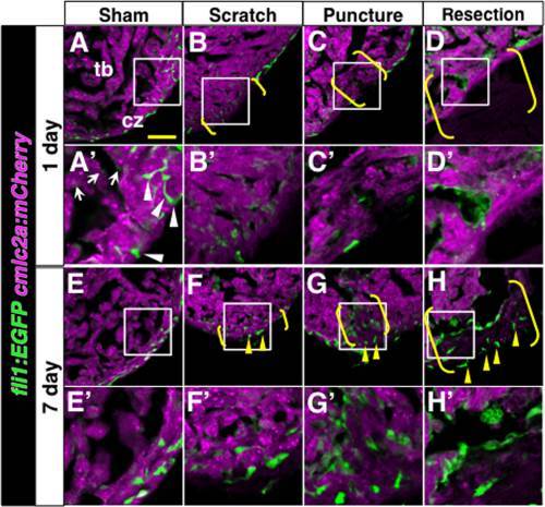
Analysis of endothelial cells after different damages to the heart. The fli1:EGFP signal (green, endothelial cells) and the cmlc2a:mCherry signal (magenta, cardiomyocytes) at 1 day (A–D, A′–D′) and 7 days (E–H, E′–H′) after various injuries. A′–H′ show close-up images of boxed areas in A–H. White arrowheads and arrows in A′ point to thick and thinner fli1:EGFP signals, respectively. For simplicity, only A′ is labeled. Yellow arrowheads point to the fli1:EGFP signals at the surface of injury sites. Brackets indicate the injury sites. Scale bar = 50 µm. cz, compact zone; tb, trabecular zone. |
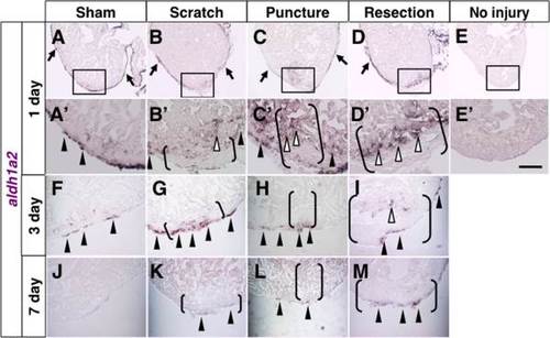
Expression of aldh1a2 after various injuries. A–D, A′–D′, F–M: In situ hybridization of aldh1a2 at 1 day (A–D, A′–D′), 3 days (F–I), and 7 days (J–M) after various injuries. E: In situ hybridization of aldh1a2 without injury. A′–E′ show close up images of boxed areas in A–E. A, A′, F: Arrowheads and arrows point to evident signals in the epicardium at the apex and away from the apex, respectively. B–D, B′–D′, G–I, K–M: Arrowheads and arrows point to evident signals in the epicardium around injury sites and away from injury sites, respectively. Open arrowheads point to endocardial signals. Brackets indicate the injury sites. Scale bar = 50 µm. |
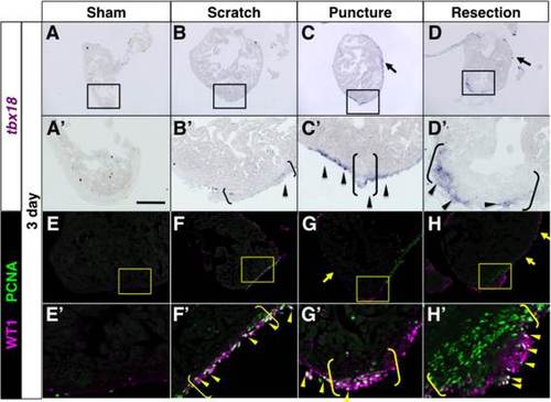
Induction of tbx18 and WT1 in the epicardium after various injuries. In situ hybridization of tbx18 (A–D, A′–D′) and immunofluorescence of WT1 (magenta) + PCNA (green) (E–H, E′–H′) 3 days after various injuries. In A–D′, arrowheads and arrows point to signals around and away from injury sites, respectively. In E–H′, arrowheads point to proliferating epicardial cells, and arrows point to WT1 signal away from injury sites. Brackets indicate the injury sites. Scale bar = 50 µm. |