
- Title
-
Overexpression of miR-19b Impairs Cardiac Development in Zebrafish by Targeting ctnnb1
- Authors
- Li, M., Hu, X., Zhu, J., Zhu, C., Zhu, S., Liu, X., Xu, J., Han, S., Yu, Z.
- Source
- Full text @ Cell Physiol. Biochem.
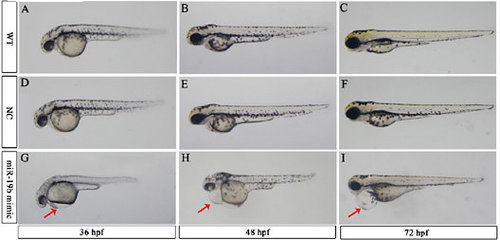
Overexpression of miR-19b leads to embryonic defects in zebrafish developmental morphology. Representative images of WT (A, B and C), NC (D, E and F), and mir-19b mimic-injected embryos (G, H and I) are shown. MiR-19b-injected embryos displayed severe pericardial edema. All images are at a magnification of 3.2 times. Red arrows: pericardial edema. |
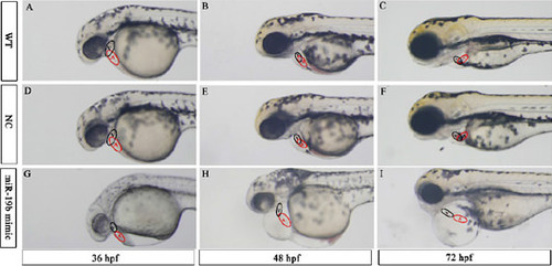
Overexpression of miR-19b leads to embryonic defects in zebrafish cardiac morphology. Representative images (6.3 times magnification) of the lateral view of WT (A, B and C), NC (D, E and F), and mir-19b mimic injected embryos (G, H, and I) are shown. In WT and NC embryos, the ventricle and atrium largely overlap with each other; however, the ventricles of mir-19b mimic-injected embryos were positioned anterior to the atrium with little overlap. Instead of the normal looped and S-shaped hearts observed in the control group, the heart chambers of miR-19b mimic-injected embryos were string-like and elongated. V: ventricle; A: atrium. |

Overexpression of miR-19b alters the expression of cardiac chamber marker genes. Representative images (8 times magnification) of 48 hpf embryos showing the expression of vmhc (A, B, C and D), amhc (E, F, G and H) and cmlc2 (I, J, K and L) by in situ hybridization are shown. After the heart tube has formed, leftward movement of the heart (jogging) is followed by rightward bending of the ventricle (D-looping) in WT embryos (A, E and I) and NC-injected embryos (B, F and J). In contrast, miR-19b mimic-injected embryos exhibited multiple LR defects, including reversed looping (L-looping; D, H and L) and no looping (C, G and K). In the bottom right-hand corner, the number of embryos examined with the representative expression/total number of embryo are presented. Black dashed lines: the midline of the body axis; red arrow: ventricle; blue arrow: atrium. EXPRESSION / LABELING:
|
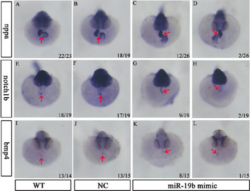
Overexpression of miR-19b has no effect on the expression of atrioventricular canal marker genes. Representative images (8 times magnification) of 48 hpf embryos showing the expression of nppa (A, B, C and D), notch1b (E, F, G and H) and bmp4 (I, J, K and L), as assessed using in situ hybridization, are shown. MiR-19b mimic-injected embryos (C, D, G, H, K and L) exhibited no significant difference in expression level and scope, except for changes in expression position due to LR defects when compared with WT embryos (A, E and I) and NC-injected embryos (B, F and J). In the bottom right-hand corner of each image, the number of embryos examined with the representative expression/total number of embryos are presented. Red arrow: atrioventricular canal. EXPRESSION / LABELING:
|
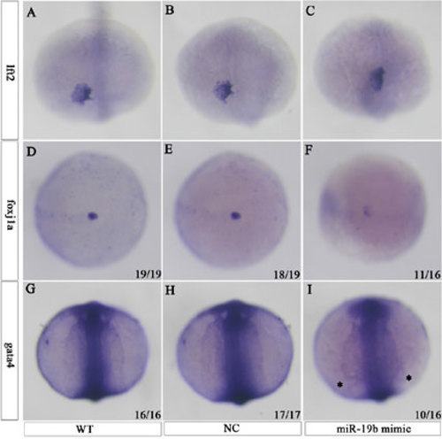
Overexpression of miR-19b altered lft2, foxj1a and gata4 expression. Representative images of lft2 expression in the cardiac field are shown using a dorsal view of 22-so-mite-stage embryos, with the anterior end at the top. The expression position of lft2 was altered in miR-19b mimic-injected embryos (C), compared with WT (A) and NC-injected (B) embryos. Representative images of foxj1a expression in DFCs are also shown. Foxj1a expression in DFCs was severely downregulated in miR-19b-overexpressing zebrafish embryos (F), compared with WT (D) and NC-injected (E) embryos. Representative images of gata4 expression are presented. Gata4 expression was restricted to the rostral Anterior lateral plate mesoderm (ALPM) in WT embryos (G) and in embryos injected with NC (H); however, its expression was expanded caudally and laterally in embryos injected with miR-19b (I). The asterisk indicates alteration of gata4 expression. EXPRESSION / LABELING:
|

MiR-19b directly targets ctnnb1. A representative image of ctnb1 expression in 48 hpf embryos determined by in situ hybridization is shown (A). The expression in the miR-19b mimic-injected embryos was decreased when compared with WT and NC-injected embryos. Similarly, real-time PCR showed the expression level of ctnnb1 at 24 and 48 hpf was significantly downregulated in the mir-19b mimic-injected embryos. A schematic of the luciferase vector-expressing ctnnb1 32UTR construct is shown (C). Luciferase assay with ctnnb1 32UTR of WT, NC and miR-19b mimic-injected embryos is presented (D). ** P < 0.01; *** P < 0.001. |

Treatment with LiCl partly rescues cardiac defects caused by overexpression of miR-19b. Representative images of cardiac morphology (A, C, E, G and I) and cmlc2 in situ hybridization (B, D, F, H and J) are shown in 48 hpf zebrafish embryos. Mir-19b mimic-injected embryos displayed severe pericardial edema (G) and looping defects (H), which were rescued by treatment with 0.15 μm LiCl immediately after miR-19b injection (I and J). In contrast, cardiac morphology and looping were normal in WT (A and B), NC-injected (C and D) and 0.15 μm LiCl-treated embryos (E and F). Red arrow: ventricle; blue arrow: atrium; yellow arrow: pericardial edema. EXPRESSION / LABELING:
|