
- Title
-
Pulses of Notch activation synchronise oscillating somite cells and entrain the zebrafish segmentation clock
- Authors
- Soza-Ried, C., Oztürk, E., Ish-Horowicz, D., Lewis, J.
- Source
- Full text @ Development

Time course of deltaC expression in heat-shocked Tg(hsp70l:dlc) transgenic embryos. (A-D) Embryos at 14-16hpf were heat-shocked at 38.5°C either once, for 15min, with recovery at 28.5°C (A) or repeatedly (B-D), with recovery at 23°C between heat-shocks. (A) Whole mounts stained by ISH with a probe for deltaC mRNA, at different times after the single heat-shock, along with wild-type controls either heat-shocked (wt hs) or not (wt). ne7 for each time point. (B) Scheme of the repetitive heat-shock treatment. (C,D) Samples were fixed for FISH with deltaC riboprobe before and after each heat-shock; fluorescence was measured by confocal microscopy as an average over the three boxed regions (30×30μm2) marked in panel Ci. (C) Specimens before and after the first (i,ii) and second (iii,iv) heat-shocks. (D) Measurements for embryos subjected to up to five heat-shocks. Values are means of n embryos fixed before and after the end of each heat-shock, where (nbefore, nafter)=(9, 10), (4, 4), (7, 4), (5, 3) and (7, 7) for the successive heat-shocks. Error bars show s.e.m. Asterisks indicate statistical significance of the difference between measurements before and after the given heat shock: *Pd0.06; **P<0.001; one-tailed t-test. EXPRESSION / LABELING:
|

tg+;dlC+ embryos subjected to a single heat-shock, compared with the wild-type and dlc/ non-transgenic controls. Transgenic tg+;dlc+ embryos at the 14-somite stage were given a single 15-min heat-shock, left to develop at 28.5°C until 48hpf, and then fixed and stained as whole mounts by ISH for the somite boundary marker cb1045. (A) Scheme of the heat-shock treatment. (B) Control heat-shocked wild-type embryo. (C) Control non-heat-shocked tgdlc (dlctw212b/tw212b, beamter) embryo. (D-F) Heat-shocked tg+;dlc+ embryos. Arrows indicate disrupted (F) or abnormally small (E) or large (D) segments. |

Rescue of segmentation by artificial pulses of deltaC expression in dlctw212b/tw212b embryos. Transgenic tg+;dlc embryos (lacking endogenous functional DeltaC) at the 11-12 somite stage were heat-shocked one, two, three or four times, or not at all, left to develop at 28.5°C until 48hpf, and stained as in Fig. 2. Recovery periods between heat-shocks were 25min at 23°C. (A) Scheme of the repetitive heat-shock treatments. (B-G) Typical results for each number of heat-shocks. Orange asterisks mark rescued somite boundaries. Yellow arrowhead in C marks the 17th somite boundary. Note that multiple heat-shocks increase the number of rescued somites: a single heat-shock produced 4.1±0.3 (mean±s.e.m.), two heat-shocks produced 4.7±0.4, three heat-shocks produced 5.0±0.3 and four heat-shocks produced 7.0±0.25. A wild-type (tgdlc+) embryo subjected to a series of four heat-shocks is included as a control; it shows normal segmentation (C). Each photo is representative of n embryos examined, where n=8 for the heat-shocked wild type, and n=20, 21, 22, 22 and 29 for the tg+;dlc embryos subjected to zero, one, two, three and four heat-shocks, respectively. |

Somite widths can be modified by varying the interval between heat-shocks. Batches of tg+;dlc embryos were taken at the 3-somite stage and heat-shocked four times as in Fig. 3, but with different recovery times, R, between one heat-shock and the next for the different batches. (A) Scheme of heat-shock treatment. (B-F) Typical results for different heat-shock repetition cycle times, from 15+15min (i.e. 15min at 38.5°C followed by 15min at 23°C) to 15+65min. (B) A control (tgdlc+) wild-type embryo heat-shocked four times on a 15+25min schedule. (C-F) tg+;dlc embryos subjected to various different heat-shock regimes as indicated. Repetitive heat-shock rescued somite boundary formation to a sufficient extent in almost every case to allow measurement of somite width. (B2-F2) Detailed views of the region measured for each batch, corresponding to the sixth to the ninth somite (red boxes in B-F). White arrowheads in D2 mark fragments of somite boundary in the middle of giant somites. (G) Histogram of mean somite width (defined as distance from the vertex of one somite-boundary chevron to the next, shown by red line in B-F) for the measured region, as a function of heat-shock repetition cycle time. Note that embryos with heat-shock repetition cycle times of 15+30min to 15+60min produce bigger somites than those observed in the non-heat shocked controls (ctrl no hs) in the evaluated region, with reversion to a size only slightly greater than control at 15+65min. *P<0.05; **P<0.005, two-tailed t-test. EXPRESSION / LABELING:
|

Rescued somite boundary formation correlates with rescued coordination of her1 oscillation. Mutant tg+;dlc embryos were taken at the 12-somite stage, given two standard 38.5°C heat-shocks separated by 25min at 23°C, and then maintained at 23°C for varying lengths of time before fixation. (A) Scheme of the treatment. (B) Wild-type embryo stained by FISH with her1 riboprobe, indicating the regions chosen for measurement (30×30µm2 yellow boxes) for all specimens in this experiment. Scale bar (vertical): 90μm. Fluorescence intensity was measured photometrically from z-stack projections. As an indicator of coordinated her1 oscillation, we took the ratio of the level of her1 in the extreme posterior box to the levels in the two symmetrically placed more anterior (mid-PSM) boxes. (C) Graph of this ratio as a function of time after the end of the second heat-shock. Two repetitions of the whole experiment are shown (brown and red lines), along with corresponding measurements for control tgdlc embryos (blue line). Each data point is an average over at least three, and on average 5.4, specimens; error bars show s.e.m. Both repetitions of the heat-shock experiment show similar clear oscillations, with period of <40min, as expected for the segmentation clock at 23°C (Schroter et al., 2008). By contrast, the control series of deltaC/ embryos without heat-shock shows only small fluctuations. The measurements for control tgdlc+ embryos are at the left-hand side of the plot, and assigned to cycling phase I, II or III [nomenclature of Pourquie and Tam (Pourquie and am, 2001)]. EXPRESSION / LABELING:
|

Time course of DeltaC protein expression in tg+;dlC+ transgenic embryos after a single heat-shock. 15 hpf embryos were heat-shocked at 38.5 ºC for 15 minutes and left to recover at 23ÚC for varying lengths of time before fixation. (A) Scheme of the treatment. (B) Flat-mounted embryo, dorsal view; white box shows region of posterior PSM selected for analysis. (C) Enlargements of boxed region marked in B, for a series of embryos fixed at the indicated times and immunostained for DeltaC (green) and beta-catenin (red, marking plasma membrane); confocal imaging. Levels of cell-surface-associated DeltaC have begun to increase at 15+15 minutes, reach a peak at 15+25 minutes, and have declined markedly by 15+30 minutes. Pictures are representative of 5 transgenic embryos analysed by confocal microscopy at each time point. |
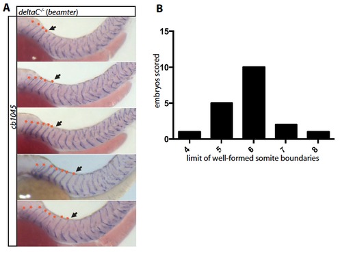
(A) Examples of dlctw212b/tw212b (tg-;dlc-, beamter) embryos, stained by ISH with cb1045 at 48hpf. Asterisks mark well-formed boundaries; arrows mark the posterior limit of segmentation. (B) Histogram showing the frequency with which different numbers of somite boundaries are formed anteriorly before segmentation breaks down. Note that the numbers of regular segments on the two sides of the embryo do not always match. The histogram shows data for one side of the embryo. |

Responses of tg+;dlc- embryos to a single standard heat-shock, showing the range of variation in the number of rescued somite boundaries. (A) Scheme of the experiment. (B) Results. Details of staining etc. as in Fig. 3. |
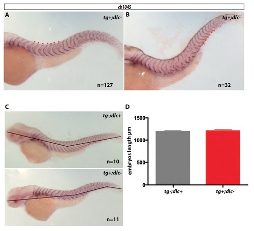
Responses of tg+;dlc- embryos to a series of 4 standard heat-shocks (15+25 minutes), delivered (A) at the 3-somite stage and (B) at the 13-15 somite stage, with fixation at 48hpf. Within each class, all embryos (n=127 for A, n=32 for B) showed a similar distribution of rescued somites – anterior for A, about 12 somite-widths further posterior for B – as illustrated by the photographs. In each case, the rescue of segmentation begins with a delay of 3 to 5 segmentation clock cycles relative to the time of heat-shock. Asterisks mark rescued somite boundaries. Details of staining etc. as in Fig. 3. (C) Measurement of whole tg-;dlC+ and tg+dlC- (15+60 minutes treatment) embryos. (D) Graph of embryo length measurements. Note that heat shocked tg-;dlC+ (n=10) and tg+dlC- (n=11) embryos have similar body lengths. |
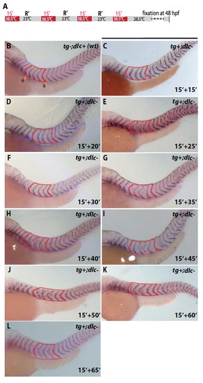
Somite width can be modified by varying the interval between heat-shocks: typical results for each of the intervals studied. (A) diagram of the heat-shocking regime. (B) Wildtype control. (C-L) Examples from batches of tg+;dlc-embryos taken at the 3-somite stage and heat-shocked 4 times, but with different recovery times, R, between one heat-shock and the next, as indicated. The red box in each case outlines the set of 4 somites whose width was measured. See Fig. 4 legend for details. |

PSM length in heat-shocked tg+;dlc- embryos is the same as in heat-shocked wildtype. (A) tg+;dlembryos, along with wildtype controls, were taken at the 13 somite stage, given a series of heat-shocks and then stained by double FISH for mespb mRNA (red channel) and deltaC mRNA (green channel). The distance from the posterior tip of the embryo to the middle of the mespb expression domain (asterisk) was measured (yellow line). (B) Graph of length measurements for tg-;dlC+ (n=5) and tg+;dlC- (n=5) embryos after 3 standard heat-shocks (15+25 minutes), fixed 25 minutes after the end of the last heat shock. The values for wildtype and mutant were respectively 364 ± 4 μm and 360 ± 7 μm (mean ± SEM). (C) Graph of length measurements for tg-;dlC+ (n=6) and tg+;dlC- (n=5) embryos after 4 heat-shocks repeated with periodicity 15 + 65 minutes, fixed 65 minutes after the end of the last heat shock. The values for wildtype and mutant were respectively 328 ± 11.6 μm and 335 ± 17 μm (mean ± SEM). The differences in B and C are not statistically significant (two-tailed t-test). |