
- Title
-
Targeted transgene integration overcomes variability of position effects in zebrafish
- Authors
- Roberts, J.A., Miguel-Escalada, I., Slovik, K.J., Walsh, K.T., Hadzhiev, Y., Sanges, R., Stupka, E., Marsh, E.K., Balciuniene, J., Balciunas, D., and Müller, F.
- Source
- Full text @ Development
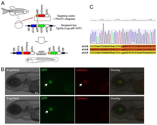
In vivo detection system for targeted integration of reporter constructs in zebrafish. (A) Schematic of PhiC31 targeted integration system. Transgenic embryos containing an attP docking site have a green lens due to the gamma-crystalline promoter driving a GFP reporter. ITR labels recognition sequences of either Tol2 or Sleeping beauty transposases used in generating the recipient transgenic lines with docking site. Injection of a circular plasmid with attB and a red reporter (targeting vector) into recipient line eggs leads to eye colour change upon PhiC31-targeted integration in larvae. (B) Detection of eye colour change upon PhiC31-mediated integration in the transgenic recipient Tg(Xla.crygc:attP-GFP)uobL6 line. Top row shows a transgenic larva with PhiC31-mediated recombination of the attB-mCherry cassette into the attP-GFP docking site. Bottom row shows transgenic sibling from the recipient line without targeted integration. Side views on cranial end of 5 dpf larvae with anterior to the left. Scale bar: 100 µm. Arrows indicate reporter expression in the lens. (C) Sequence of the tpl102 recipient docking site with germline integration of targeting construct tnnT2:attB-mRFP. Sequence shows the attR site in the crygc:attR-Red recombination product. The three lower case nucleotides denote the recombination site. |

Variability of position effects on reporter activity is sharply reduced among PhiC31 integrase-mediated transgenic lines compared with Tol2 transgenesis. (A) Schematic of the pTol2/EL161-krt4:Venus construct injected with Tol2 transposase mRNA. Below are examples of expression patterns of transgenic F1 larvae (3 dpf). TP indicates Tol2-mediated expression patterns. Numbers refer to domains of Venus expression activity (listed in Fig. 3B). (B) Schematic indicates the recombination of the targeting plasmid pattB-mCherry,Tol2/EL161-krt4:Venus into the attP docking site of the transgenic recipient line Tg(Xla.crygc:attP-GFP)uobL6. Larvae from three different founders (IF1, IF2, IF8) targeted with PhiC31 integrase are shown. Venus expression domains are labelled as in Fig. 3B. Lens activity driven by mCherry (arrowheads in bottom row) demonstrates PhiC31-mediated integration. Insert in bottom right shows brightfield image of the head region. Arrows labelled ‘ysl’ in red channel indicate auto-fluorescence of the yolk syncytial layer. Dorsal views of larvae head are shown. Scale bar: 100 μm. |
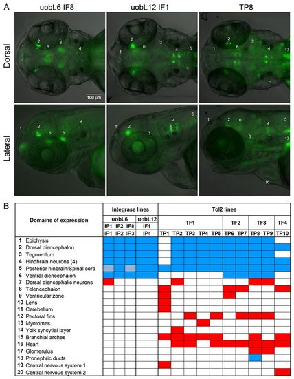
Reproducible Venus expression patterns upon targeted integration of EL161 enhancer construct demonstrate cis-regulatory function in the brain. (A) Brain-specific enhancer effect of a transgene inserted in different integration sites. Domains of Venus activity are specified in B. Dorsal (top) and lateral (bottom) views of the head of 3 dpf F1 transgenic larvae. Scale bar: 100 μm. (B) Domains of Venus reporter expression and their distribution among targeted (PhiC31 integrase) and randomly (Tol2 transposase) transgenic lines. IF, integrase-injected founders; IP, integrase-mediated expression patterns; TF, Tol2-injected founders; TP, Tol2-mediated expression patterns. Blue depicts expression domains overlapping with esrrga activity, whereas red colour indicates additional domains. Light blue indicates weak expression. |

Transient transgenesis strategy for rapid evaluation of enhancer activity upon PhiC31-mediated targeting. (A) Schematic of PhiC31-targeted integration system, designed for cis-regulatory element analyses in transient transgenic embryos. ‘-2.4 shh’, 2.4 kb upstream region of shh gene; ‘shhABC’, shh complete introns 1 and 2, with ar-A, ar-B, ar-C enhancers (Müller et al., 1999; Ertzer et al., 2007). (B) Detection of reporter activity in embryos injected with construct containing the shh cis-regulatory elements by in situ hybridisation (B) and fluorescence microscopy (C). Arrows indicate activity in shh expression domains, resulting from successful integration of the shh cis-regulatory cassette in to the attP site of uobL12, whereas arrowheads indicate the activity of the crygc promoter in uobL12 recipient line. Scale bars: 150 μm. |

Tol2 transgenic founders show variable position effects on EL161 driven reporter activity. All of the different expression patterns shown by Tol2 transgenic lines injected with pTol2/EL161-krt4: Venus construct. Tol2 patterns (TP) are numbered from 1 to 10. Dorsal and lateral views of YFP and brightfield channels are shown and annotated domains correspond to the expression domains described in Figure 3B. Larvae are protruding mouth stage (approximately 3 dpf ) with anterior to the left. Scale bar indicates 100 μm. |

PhiC31-mediated transgenic founders show highly reproducible expression patterns. Neuronal expression patterns obtained in integrase injected founders uobL6 IF1, uobL6 IF2, uobL6 IF8 and uobL12 IF1. Red lens indicates legitimate PhiC31-mediated integration into the Xla.crygc:attP-GFP cassette from either Tg(Xla.crygc:attP-GFP)uobL6 or Tg(Xla.crygc:attP-GFP)uobL12. Auto-fluorescent yolk syncytial layer (ysl) is marked by arrows in mCherry images. YFP channel shows EL161-driven expression patterns are listed in Figure 3B. Larvae are at protruding mouth stage (3 dpf ) with anterior to the left. Scale bar indicates 100 μm. |
|
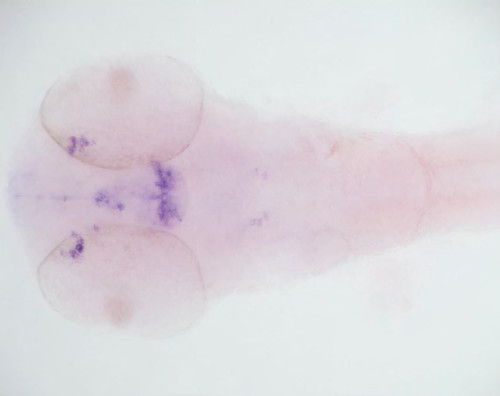
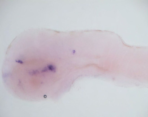
Tissue-specific activity driven by EL161 element overlaps with endogenous expression domains of endogenous esrrga gene. 60 hpf embryos probed with esrrga (left) share 5 expression domains with integrant founders from uobL6 (right) probed with Venus. Dorsal and lateral images are shown. Expression domains are labelled as listed in Fig. 3B. Note that esrrga domain 1 (epyphsis) is weakly expressed at this stage, but shows a strong expression earlier in development. Lens from bottom left embryo was removed to allow visualization of inner brain domains. Scale bar indicates 100 μm. 


|

EL161 putative enhancer from essrga locus drives a complex and dynamic neuronal pattern. Complexity and dynamic pattern of pattB-mCherry,Tol2/EL161-krt4:Venus construct integrated in uobL6 IF1 are shown from 60 hpf to 96 hpf. Red lens indicates PhiC31-mediated legitimate integration. YFP channel shows the dynamic expression pattern driven by the injected construct. Domains of expression are listed in Figure 3B. All larvae are oriented anterior to the left. Scale bar indicates 100 μm. |