
- Title
-
A unique covalent bond in basement membrane is a primordial innovation for tissue evolution
- Authors
- Fidler, A.L., Vanacore, R.M., Chetyrkin, S.V., Pedchenko, V.K., Bhave, G., Yin, V.P., Stothers, C.L., Rose, K.L., McDonald, W.H., Clark, T.A., Borza, D.B., Steele, R.E., Ivy, M.T., Hudson, J.K., and Hudson, B.G.
- Source
- Full text @ Proc. Natl. Acad. Sci. USA

NC1 hexamers were excised from animal basement membranes and analyzed by SDS/PAGE as shown in Fig. 1 A and B. The dimeric subunits, which indicate the presence of the bond, were found in nine major eumetazoan phyla. Among eight cnidarians investigated, only Hydra NC1 lacked dimeric subunits. All NC1s were immunoblotted against the rat monoclonal antibody, JK2, except for C. elegans (rabbit polyclonal; NW-154) and Drosophila (mouse monoclonal; 6G7). Black outlines indicate the locations of cropping for blot images. EXPRESSION / LABELING:
|
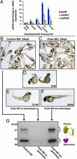
Expression of collagen IV and Pxdn during development in zebrafish and morpholino (MO) knockdown of peroxidasin in zebrafish embryos. (A) Pxdn and collagen IV expression during zebrafish embryonic development. Real-time qPCR studies were conducted to examine expression levels of Pxdn, collagen4α1, and collagen4α2. *Student t test P value < 0.03 compared with expression at 1,000 cells. Error bars = SEM. Blue, pxdn; red, col4a; black, col4a2. (B) Control and (C) Pxdn MO groups. MO-injected embryos displayed (D) general severe defects that include cardiac edema, smaller eyes, and gross trunk patterning defects (4/45), (E) partial curved trunk (21/45), or (F) normal development (20/45). (G) SDS/PAGE analysis of Pxdn MO embryonic phenotypes at 24 hpf by Western blot. Collagenase digests were normalized for total protein load by protein stain with SYPRO-Ruby (Fig. S8). |