
- Title
-
Maternal control of axial-paraxial mesoderm patterning via direct transcriptional repression in zebrafish
- Authors
- He, Y., Xu, X., Zhao, S., Ma, S., Sun, L., Liu, Z., and Luo, C.
- Source
- Full text @ Dev. Biol.

Maternal Vsx1 is essential for axial–paraxial mesoderm patterning in zebrafish. (A–C) Uninjected wild-type control embryos. (D–F) vsx1 tbMO injected embryos exhibit dorsalized phenotype with disturbed dorsal midline structure. (G–I) vsx1 overexpression elicits inhibition of axial mesoderm specification and aberrant convergence of specified paraxial mesoderm. (J–L) Injection of vsx1 sbMO has no detectable effect on development before 24 hpf. Phenotypes are shown at 24 hpf. (M) Proportion of different phenotypes in vsx1 knockdown, overexpression and rescue experiments at 24 hpf. DDMS: disorganized dorsal midline structure. SB: spina bifida. PHENOTYPE:
|
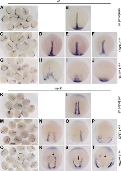
Comparison of axial and paraxial mesoderm formation among wild type, maternal Vsx1 suppressed and vsx1 overexpression embryos. (A–J) ntl marked axial mesoderm domain in uninjected wild-type control embryos (A and B), vsx1 tbMO injected embryos (C–F) and vsx1 mRNA injected embryos (G–J). (K–T) myoD marked paraxial mesoderm domain in uninjected wild-type control embryos (K and L), vsx1 tbMO injected embryos (M–P) and vsx1 mRNA injected embryos (Q–T). Note that vsx1 overexpression inhibits the convergence of paraxial mesoderm cells but has no impact on paraxial mesoderm cell specification and somite formation. Riboprobes are indicated at the top of each group of figures. All the images of single embryo are dorsal view with animal pole towards the top. EXPRESSION / LABELING:
PHENOTYPE:
|

Maternal Vsx1 is essential for promoting normal paraxial mesoderm specification. (A–F) comparison of msgn1 expression between uninjected wild type control (A–C) and maternal vsx1 tbMO injected embryos (D–F). (G–L) Comparison of tbx24 expression between uninjected wild type control (G–I) and maternal vsx1 tbMO injected embryos (J–L). Riboprobes are indicated at the bottom of each figure. (B, E, H and K) Dorsal view of embryos with animal pole towards the top. (C, F, I and L) Animal pole view of the embryos with dorsal towards the right. EXPRESSION / LABELING:
|
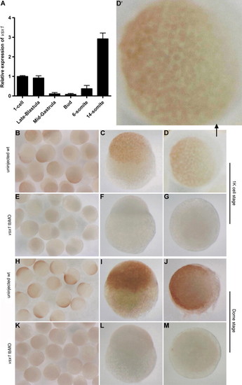
Expression pattern of vsx1 during early embryogenesis. (A) Expression profile of vsx1 mRNA during early embryogenesis. Results are expressed as mean±SEM. (B–M) Localization of maternal Vsx1 protein in uninjected wild type control and vsx1 tbMO injected embryos at 1k cell (B–G) and dome stages (H–M). (C, F, I and L) Lateral view of embryos with animal pole towards the top and dorsal towards the right. (D, D′, G, J and M) Animal pole view of the embryos with dorsal towards the right. D′ is the magnified image of D, in which one can see that Vsx1 protein is localized in the nuclei of the blastomeres. |
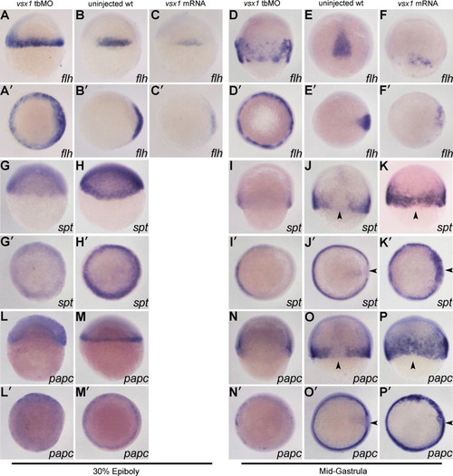
Vsx1 represses flh to preserve spt and papc expression in the ventolateral margin. The injected reagents are indicated at the top of each column. Riboprobes are indicated at the bottom of each figure. (A–P) Dorsal view of embryos with animal pole towards the top. (A′–P′) Animal pole view of the embryos with dorsal towards the right. Arrow heads indicate that the paraxial mesoderm marker is detected in the presumptive axial mesoderm region in vsx1 overexpression embryos. EXPRESSION / LABELING:
|

Injection of vsx1 tbMO has no obvious influence on the expression patterns of wnt8, bmp2b and their target vent/vox at shield stage. The injected reagents are indicated at the left side of the images. Riboprobes are indicated at the top of images. The images of single embryo are animal pole view with dorsal towards the right. EXPRESSION / LABELING:
|

Vsx1 is a transcriptional repressor. (A) Schematic representation of the Vsx1 mutants. (B) Comparison of flh promoter mediated gfp expression under the regulation of a fusion protein En–Vsx1 and a fusion protein VP16–Vsx1. Results are expressed as mean±SEM, and statistical analyses were done by unpaired t test. ***P<0.001. (F–H) En–Vsx1 fusion protein functions as a wild type Vsx1. (I–K) VP16–Vsx1 fusion protein functions as a Vsx1 antimorph. The injected reagents are indicated at the right side of the images and riboprobes are indicated at the bottom of each figure. (C, F and I) Dorsal view of phenotypes at 8–10 somite stage with head towards the top. (D, G and J) Dorsal view of embryos with animal pole towards the top. (E, H and K) Animal pole view of the embryos with dorsal towards the right. In situ hybridization of flh is shown at 30–40% epiboly stage. |
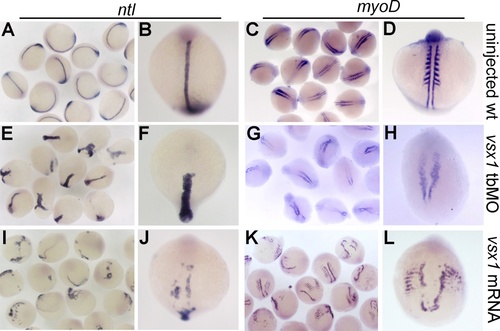
Comparison of axial and paraxial mesoderm formation in uninjected wildtype control, vsx1 knockdown and overexpression embryos at 8-10 somite stage. (A-B, E-F, I-J) Axial mesoderm domain is visualized by whole mount in situ hybridization with marker gene ntl. (C-D, G-H, K-L) Paraxial mesoderm domain is visualized by whole mount in situ hybridization with marker gene myoD. |

Vsx1 binding site 11 mediates Vsx1-dependent repression of GFP driven by flh proximal promoter. Both wild-type and mutant type of flh proximal promoter fragments drove GFP expression successfully and ubiquitously after middle blastula stage. When co-injected with vsx1 mRNA, GFP expression from the wild type promoter fragments was suppressed but from the mutant promoter fragment was kept. The types of GFP reporter sensors are shown on the top of the images and the co-injected mRNA is shown on the left of the images. |
Reprinted from Developmental Biology, 386(1), He, Y., Xu, X., Zhao, S., Ma, S., Sun, L., Liu, Z., and Luo, C., Maternal control of axial-paraxial mesoderm patterning via direct transcriptional repression in zebrafish, 96-110, Copyright (2014) with permission from Elsevier. Full text @ Dev. Biol.