
- Title
-
Toll-like receptor 9 and 21 have different ligand recognition profiles and cooperatively mediate activity of CpG-oligodeoxynucleotides in zebrafish
- Authors
- Yeh, D.W., Liu, Y.L., Lo, Y.C., Yuh, C.H., Yu, G.Y., Lo, J.F., Luo, Y., Xiang, R., and Chuang, T.H.
- Source
- Full text @ Proc. Natl. Acad. Sci. USA
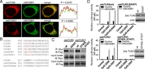
Regulation of zebTLR9 and zebTLR21 activation by UNC93B1. HEK293 cells were cotransfected with expression vectors for FLAG-tagged WT and mutant zebTLR9 and zebTLR21, Myc-tagged UNC93B1, and NF-κB luciferase reporter gene as indicated. (A) Cellular localizations of proteins were visualized by immunofluorescence staining. Colocalizations of UNC93B1 with zebTLR9 and zebTLR21 were quantified by calculating the Pearson correlation coefficient of the signaling intensities of the two proteins. (C) Protein binding was measured by immunoprecipitation followed by immunoblotting. (D) Cells were treated with 3 µM CpG-ODN, after which relative luciferase activity was measured. Data are mean ± SD (n = 3). Immunoblots shown represent expression levels of WT and acidic amino acid residue-mutated zebTLR9 and zebTLR21. (B) Sequence alignment of the juxtamembrane regions of different human and zebrafish TLRs. The acidic amino acid residues in intracellular TLR regions are shown in red. |

Antimicrobial activities of different CpG-ODNs in zebrafish. Thirty zebrafish in each group were injected i.p. with 5 × 10⁵ cfu of E. tarda with or without 1 μg of CpG-ODN as indicated. (A and B) Skin hyperemia (A) and H&E staining (B) of sections from fish on day 2 after injection with/without E. tarda. (C) Cumulative percent mortality of fish after injection with E. tarda and CpG-ODN. |

Expression of zebTLR9, zebTLR21, and zebUNC93B1 at different times during embryonic development (A) and in different tissues of adult zebrafish (B). Expression of the indicated genes was analyzed by RT-PCR. Amplification of GAPDH was performed for control of amounts of cDNA used as a template. Blots are representative RT-PCR analyses of three independent experiments. |

Intracelullar activation and localization of zebTLR9 and zebTLR21. (A) HEK293 cells were cotransfected with expression vector for zebTLR9 (Left) and zebTLR21 (Right) and NF-κB luciferase reporter gene. These cells were pretreated with 2 μM chloroquine, and then treated with 0.2 μg/mL flagellin and 3 μM CpG-ODN. Relative luciferase activities were determined. Data are mean ± SD (n = 3). *P < 0.05 vs. cells without chloroquine treatment. (B) HEK293 cells were transfected with expression vector for zebTLR9 and zebTLR21. Expression of zebTLR9 and zebTLR21 was visualized by immunofluorescence staining using anti- FLAG M2 mAb, followed by an Alexa Fluor 488-labeled anti-mouse antibody. Cellular organelles were stained by anti-Lamp1, anti-AIF, anti-EEA1, anti-RCAS1, and anti-calnexin antibodies, followed by an Alexa Fluor 594-labeled secondary antibody. Nuclei were stained by DAPI. Representative merged confocal images are shown for the cellular localization of zebTLR9 and zebTLR21. Green indicates zebTLR9 or zebTLR21; red, organelle markers; blue, nuclei stained with DAPI. |