
- Title
-
Mediator subunit 12 coordinates intrinsic and extrinsic control of epithalamic development
- Authors
- Wu, S.Y., de Borsetti, N.H., Bain, E.J., Bulow, C.R., and Gamse, J.T.
- Source
- Full text @ Dev. Biol.

The med12/kgt mutant lacks the parapineal organ. (A-B′) Compared to WT (white arrow in A; dotted circle in B), med12 mutants showed no parapineal formation at 2 or 3 dpf (A′, B′) as revealed by the Tg[foxd3:GFP] transgene (used in all images to label the pineal complex unless indicated otherwise). (C-E′) Expression of parapineal markers in med12 mutants compared to WT embryos revealed by WISH (otx5 and gfi1.2 at 4 dpf; sox1a at 32 hpf). No parapineal markers were expressed in med12 mutants (C′, D′, E′), in contrast to WT embryos (C, D, E; black arrows). |
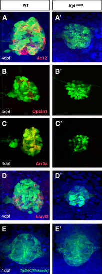
Photoreceptors and projection neurons are not maintained in the pineal complex of med12 mutants. Rod photoreceptors were absent at 4 dpf in med12 mutants, as labeled by immunofluorescence for 4c12 (A, A′) and Opsin1 (B, B′). Cone cells are also absent in med12 mutants at 4 dpf, as shown by Arr3a staining (C, C′). Elavl3 staining showed a reduction in the number of the projection neurons at 4 dpf (D, D′). med12 mutants and WT showed comparable expression of the TgBAC[flh:kaede] transgene (E, E′), which labels the pineal complex at 1 dpf. Cell nuclei are stained with ToPro3 (blue). |
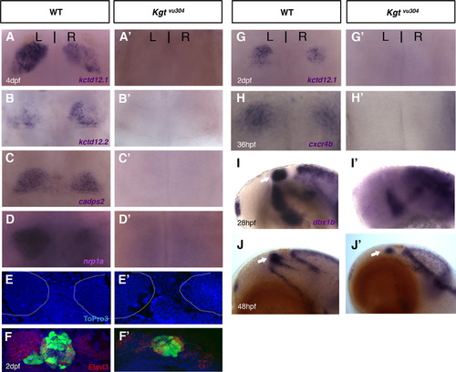
The med12/kgt mutant shows defects in habenular development. The habenular differentiation markers, kctd12.1, kctd12.2, nrp1a, and cadps2 were all absent in med12 mutants at 4 dpf (A-D′) and 2 dpf (G, G′), while the habenular cells were still present in the dorsal diencephalon in med12 mutants as shown by ToPro3 nuclear staining at 4 dpf (E, E′; habenular nuclei were outlined in white; the dark shadow on the left habenula in E′ is due to an overlying pigment cell). Fewer Elavl3-expressing neurons were observed (F, F′; at 2 dpf) and the expression of cxcr4b was significantly reduced in med12 mutants when compared to WT (H, H′; at 36 hpf). In med12 mutants, the presumptive habenular progenitor marker, dbx1b, was absent early (lateral views, at 28 hpf; I, I′) but reappeared in later stages (at 48 hpf, J, J′). |
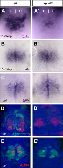
Loss of Med12 activity leads to reduction in tbx2b expression and FGF signaling activation. Compared to WT, the expression of tbx2b was decreased in med12 mutants at 14 hpf (A, A′); while the expression of flh remained unchanged (B, B′). The activation of FGF signaling was also disrupted in med12 mutants at 1 dpf as shown by Tg[dusp6:d2eGFP] transgene expression (D, D′; projection neurons labeled in red by Tg[HuC:Kaede]) and dpERK staining (E, E′), while the expression of the ligand fgf8a was unaffected (C, C′). EXPRESSION / LABELING:
|

Proper expression of photoreceptor transcription factors requires Med12. Med12 mutant at 1 dpf showed reduced levels of expression of the rod photoreceptor specification gene, nr2e3 (A, A′). The expression levels of neurod (B, B′) and the expression domain of crx (C, C′) were also reduced in med12 mutants. EXPRESSION / LABELING:
|
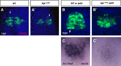
The parapineal and habenular phenotypes in med12/kgt mutants are not due to elevated apoptosis. TUNEL labeling shows no difference in the number of apoptotic cells between WT and med12 mutants in the dorsal diencephalon at 1 dpf (A, A′). The med12;tp53 double mutants lack parapineal cells (B, B′; 3 dpf) and show reduced expression of tbx2b (C, C′; 13 hpf). EXPRESSION / LABELING:
|

Timely activation of tbx2b expression is crucial for the parapineal specification. At 14 s/16 hpf stage, the expression of tbx2b was reduced only in med12 mutants (B), but not in tbx2b/lor mutants (C). Conversely, compared to WT at 1 dpf, the expression of tbx2b in med12 mutants was not significantly changed (D, E); while decreased in tbx2b/lor mutants (F). Formation of the parapineal organ is similar to WT in tbx2b/lor mutants (G; gfi1.2 at 4 dpf). EXPRESSION / LABELING:
|
Reprinted from Developmental Biology, 385(1), Wu, S.Y., de Borsetti, N.H., Bain, E.J., Bulow, C.R., and Gamse, J.T., Mediator subunit 12 coordinates intrinsic and extrinsic control of epithalamic development, 13-22, Copyright (2014) with permission from Elsevier. Full text @ Dev. Biol.