
- Title
-
Dynamic expression of neurexophilin1 during zebrafish embryonic development
- Authors
- Thomas-Jinu, S., and Houart, C.
- Source
- Full text @ Gene Expr. Patterns
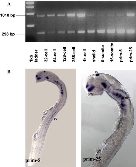
Zebrafish nxph1 expression during embryonic development. (A) RT-PCR showing the presence of maternal nxph1 in zebrafish embryos. Lane 2–11: cDNA amplified from total RNA of 32-cell, 64-cell, 128-cell, 256 cell, 1000-cell, shield, 5-somite, 15-somite, prim-5 and prim-25 stage embryos, respectively. (B) Lateral view: anterior to the left of embryos at prim-5 and prim-25. nxph1 is expressed in discrete clusters in the forebrain, midbrain, hindbrain and spinal cord. Abbreviations: t, telencephalon; pOA, post-optic area; e, epiphysis; tg, tegmentum; rh, rhombomeres; sc, spinal cord. Scale bar = 100 µm. |

Dynamic expression of nxph1 during zebrafish development. (I) Lateral (A–C) and dorsal (D–F) view of 24 hpf (A, D), 36 hpf (B, E) and 5 dpf (C, F) embryos showing nxph1 expression (blue). Black arrowhead indicates anterior and post-optic commissure and asterisk indicates hindbrain radial glia in C, F. Abbreviations: t, telencephalon; pOA, post-optic area; e; epiphysis; tg, tegmentum. (II) Transverse section showing nxph1 expression in telencephalon (t) and pre-optic area (poa) (G, H) at 24 hpf and 36 hpf. Transverse section through the diencephalon (I, white arrowhead indicates the pineal gland). Dorsal view of hindbrain (J), lateral view (K) and transverse section (L) of the spinal cord at 24 hpf. (III) Lateral (M) and dorsal (N) view of brain and transverse section of spinal cord (O) of nxph1 (red) at 36 hpf. Scale bar = 40 µm. |
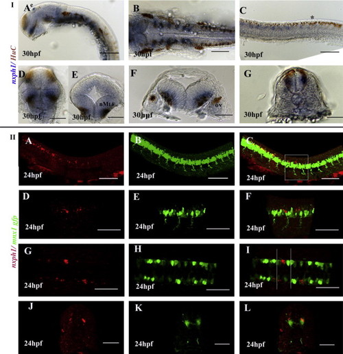
nxph1 is expressed in neurons, mostly interneurons. (I) Anti-HuC staining for post-mitotic neurons in 30 hpf embryos stained for nxph1 (A–G). Lateral view of zebrafish brain (A), frontal view of forebrain (D), transverse view of midbrain (E), dorsal (B) and transverse (F) view of hindbrain, lateral (C) and transverse (G) view of spinal cord at 36 hpf. Abbreviations: nMLF, nuclei of the media longitudinal fascicle); ov, otic vescicle; e, epiphysis. Scale bar: A–C (40 µm) and D–G (20 µm). (II) Lateral (A–F), dorsal view (G–I) and transverse section of spinal cord (J–L) of Tg(mnx1:GFP) zebrafish at 24 hpf, anterior to the left. Scale bar: A–C, J–L (40 µm) and D–I (20 ¼m). nxph1 is expressed ventral (blue) to the Rohon-Beard sensory neurons () stained by HuC (I) and dorsal (red) to the motor neurons marked by Tg(mnx1:GFP)(II), thus indicating its expression in the interneurons. |
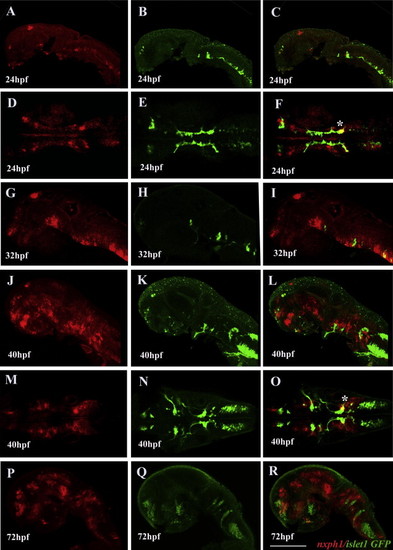
Motor neurons do not express nxph1 except for a small cluster of facial neurons. Lateral (A–C, G–L, P–R) and dorsal view (D–F, M–O) of zebrafish brain; anterior to the left. nxph1 expression in Tg(isl1: GFP) at 24 hpf (A–F), 32 hpf (G–I), 40 hpf (J–O) and 72 hpf (P–R). () indicates co-expresssion in nVII facial motor neurons. Scale bar = 50 µm. EXPRESSION / LABELING:
|
Reprinted from Gene expression patterns : GEP, 13(8), Thomas-Jinu, S., and Houart, C., Dynamic expression of neurexophilin1 during zebrafish embryonic development, 395-401, Copyright (2013) with permission from Elsevier. Full text @ Gene Expr. Patterns