
- Title
-
Sequential effects of spadetail, one-eyed pinhead and no tail on midline convergence of nephric primordia during zebrafish embryogenesis
- Authors
- Huang, C.J., Wilson, V., Pennings, S., MacRae, C.A., and Mullins, J.
- Source
- Full text @ Dev. Biol.
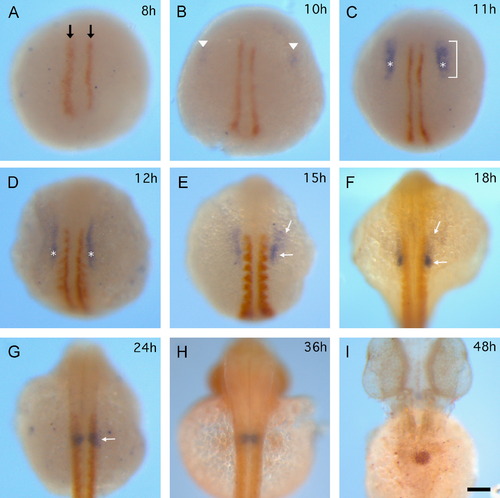
Morphogenetic changes of PGP during 8-48 hpf development. Dynamic wt1a expression (blue) in relation to somite morphogenesis (myoD expression, orange-brown) at 8, 10, 11, 12, 15, 18, 24 hpf is shown in (A)-(G). (A) MyoD expression labels adaxial cells at 8 hpf (black arrow). (B) Initial wt1a expression (arrowhead) locates bilaterally at a distance from the adaxial cells. ((C) and (D)) Wt1a expression pattern expands along the (A)-(P) dimension (bracket). Wt1a expression crosses on left-right axis at third somite were the measure points for inter distance of PGP (asterisks). ((E) and (F)) White arrows indicate faint (anterior side) and dense (posterior side) signal in wt1a-positive cells. (G) The faint signal is almost unseen and wt1a-positive cells become compact round shape (white arrow). (H) The bilateral PGP (blue) adjacent to each other at 36 hpf, myoD and mibp2 were stained orange-brown. (I) A single fuse PGP (orange-brown) appears in midline at 48 hpf. All images are dorsal view and at the same magnification. The scale bar in H indicates 100 µm. EXPRESSION / LABELING:
|

Varied PGP phenotypes in Zoep-/- embryos. (A) Wt1a (blue) and myoD (orange) double in situ shows normal PG pattern at 12 hpf (6-somite) in Zoep-/-embryos. Both normal (B) and abnormal (D) PG (arrows) phenotype is observed at 24 hpf. Mild (C) and severe PG (arrows) midline convergence defect is observed at 48 hpf. All images are dorsal view anterior to the top and at the same magnification. The scale bar in E indicates 100 µm. (F) Zoep-/- inter distance of PGP (red squres) is compare to WT on it2s plot at 12 (n=40, P=0.15), 16 (n=24), 24 (n=22), 36 (n=34), 48 (n=29) hpf. P<0.05, *P<0.0005, ***Error bars represent SD. EXPRESSION / LABELING:
|

PGP differentiation in Zoep-/- embryos. Whole-mount in situ hybrydization of wt1a ((A), (D), (G)), podocin ((B), (E), (H)) or nephrin ((C), (F), (I)) was stained blue in WT ((A)-(C)) and Zoep-/- ((D)-(I)) embryos at 36 hpf. The numbers (µm) on Zoep-/- embryos ((D)-(I)) indicate the inter distance (µm) of PGP. All images are dorsal view anterior to the top and at the same magnification. EXPRESSION / LABELING:
|
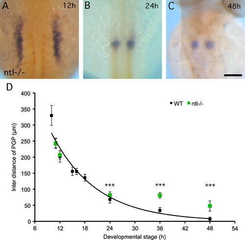
PGP midline convergence phenotypes in ntl-/- embryos. ((A)-(E)) Whole-mount in situ hybrydization of wt1a and myoD was stained blue and orange, respectively in 12 (A), 24 (B) and 48 hpf (C) ntl-/- embryos. The scale bar indicates 100 µm; ((A)-(C)) are at the same magnification. All images are dorsal view anterior to the top. (D) Quantification of convergence morphogenetic movements in ntl-/- embryos and comparison with WT. The inter distance of PGP was measured at 11 (n=20, P=0.68), 12 (n=17, P=0.27), 24 (n=23,), 36 (n=29), 48 (n=20) hpf. The inter distance of PGP is significant difference between ntl-/- and WT at 24, 36 and 48 hpf (***P<0.0005). Error bars represent SD. EXPRESSION / LABELING:
|

PGP midline convergence phenotypes in spt-/-embryos. ((A)-(C)) Whole-mount in situ hybrydization of wt1a and myoD was stained blue and orange, respectively in 12 (A), 24 (B) and 48 hpf (C) spt-/- embryos. The scale bar indicates 100 µm; ((A)-(C)) are at the same magnification. All images are dorsal view anterior to the top. (D) Quantification of convergence morphogenetic movements in spt-/- embryos and comparison with WT. The inter distance of PGP was measured at 11 (n=28), 12 (n=32), 15 (n=31), 24 (n=9), 48 (n=15) hpf. The P values of t-test for all 5 stages are smaller than 0.005. The curve fitting (pink) and R-squared value are displayed on the right-hand corner. A turning point (arrow) to divergence movement at 33.5 hpf is generated from an approximation of the stage that has the minimum inter distance of PGP. Error bars represent SD. EXPRESSION / LABELING:
PHENOTYPE:
|
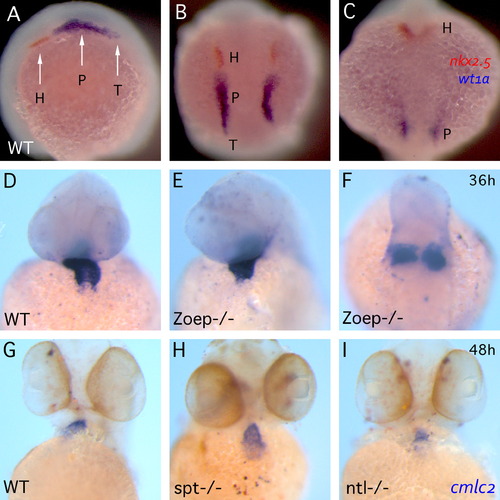
PGP and cardiac midline convergence. ((A)-(C)) Transiency of PGP neighboring tissue. Double in situ of nkx2.5 (orange) and wt1a (blue) at 12 ((A)-(B)) and 18 hpf (C). (A) The image is lateral view anterior to the right. ((B)-(C)) are dorsal view anterior to the top and same magnification. (H) heart primordium; (P) pronephric glomerular primordium; (T) tubule primordium. ((D)-(I)) Whole-mount in situ hybridization of cmlc2 demonstrating cardiac morphogenetic phenotypes at 36 ((D)-(F)) and 48 hpf ((G)-(I)) in WT ((D) and (G)), Zoep-/- ((E) and (F)), spt-/- (H) and ntl-/- (I) embryos. ((D)-(I)) The images are same magnification and anterior to the top; ((A) and (B), (D)-(F)) are ventral view and (C) is dorsal view. EXPRESSION / LABELING:
PHENOTYPE:
|

Proximal convoluted tubules and PGP differentiation. Double in situ of pod and slc20a1 ((A)-(C) and (I)) or nph and slc20a1 ((D)-(H)) in WT ((A) and (D)), ntl morphant ((B) and (E)), spt morphant ((C) and (F)), Zoep-/- embryos ((G)-(I)) at 48 hpf. Arrows indicate proximal convolute tubules and arrowheads indicate PGP. All images are dorsal view anterior to the top and same magnification. EXPRESSION / LABELING:
|
Reprinted from Developmental Biology, 384(2), Huang, C.J., Wilson, V., Pennings, S., MacRae, C.A., and Mullins, J., Sequential effects of spadetail, one-eyed pinhead and no tail on midline convergence of nephric primordia during zebrafish embryogenesis, 290-300, Copyright (2013) with permission from Elsevier. Full text @ Dev. Biol.