
- Title
-
Dermal fin rays and scales derive from mesoderm, not neural crest
- Authors
- Lee, R., Thiery, J.P., and Carney, T.J.
- Source
- Full text @ Curr. Biol.
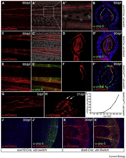
Fin osteoblasts derive from late immigrating paraxial mesoderm. (A, B) Adult (90 dpf) sox10:Cre; ubi:switch transgenics showing derivatives in the fin derived from neural crest and sox10 expressing cells. Lateral view of wholemount fin, immunostained to detect mCherry, indicates labelled cells running along the fin rays (A). Superimposing on a brightfield image demonstrates that these run within the lepidotrichia (A′). Magnified view of part of A′ highlights the position within the ray and also mCherry-positive melanophores (arrowheads; A′′). Transverse cryosection of a sox10:Cre; ubi:switch fin immunostained for mCherry (red) and zns-5 (green) and counterstained with DAPI (blue). mCherry positive cells are concentrated in two bundles within the fin ray and are zns-5 negative (B). (C–F′) Adult (90 dpf) fins of tbx6:Cre; ubi:switch (C–D′) and tbx6:CreERt2; ubi:switch (E–F′) imaged as lateral wholemounts (C–C&prime& E–E′) or as transverse cryosections (D–D&prime& F–F′). Fins have been immunostained for mCherry (red; C–F′) and zns-5 (green; D′,E′,F′) and counterstained with DAPI (blue; D′,F′). (C) is also shown superimposed on the Nomarski image (C′). In both lines, osteoblasts are mCherry positive. (G–H) Lateral images of the trunk/tail (G) and dorsal medial fin (H) of 5 dpf (G) and 21 dpf (H) tbx6:CreERt2; ubi:switch transgenics treated with 4-hydroxytamoxifen and immunostained for mCherry. Larvae with no labelled cells in the fins at larval stages (G) often were observed to have chains of cells, aligned to forming bony rays, within the fins at 21 dpf (arrows; H). (I) Counts of mCherry-positive cells in the fins during postembryonic development. Secondary immigration was noted from 14 dpf. (J–K′) Lateral images of scales in 30 dpf sox10:Cre; ubi:switch (J–J′) and tbx6:Cre; ubi:switch (K–K′) transgenics immunostained for mCherry (red; J–K′) and zns-5 (green; J′,K′). Co-labelling is only seen in the tbx6:Cre; ubi:switch line. |
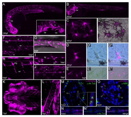
The sox10:cre; ubi:switch line permanently labels all neural crest derivatives A-L: Images of sox10:cre; ubi:switch embryos and larvae at 24hpf (A), 48hpf (B-D′′), 4dpf (E-E′′), 5dpf (F-J) and 7dpf (K-L). Live confocal (A-C′, F-L) and epifluorescent (D-E′′) images showing 2 mCherry expression in magenta. All are viewed laterally, except K and L which are ventral views. Brightfield views (D′, E′′) and overlays (D′′,E′′) are shown to visualise pigmentation. Neural crest cells can be first observed expressing mCherry weakly from 24hpf (A) and mCherry levels accumulate from there onwards, such that by 48hpf there is comprehensive expression in migrating neural crest cells (B). Neural crest cells in the branchial arches (ba) are labelled (A(inset), B). This line additionally labels the sox10 expressing otic vesicle (o; A(inset)-B). All three chromatophore lineages are broadly labelled, namely black melanophores (C-C′), iridescent iridophores (D-D3) and yellow xanthophores (E- E′′, J). As melanin strongly quenches the fluorescent signal, we visualised melanophores in young or partially PTU treated embryos. Counts of melanophores in 5 larvae at 54hpf showed 96.6 ± 0.4% were visibly mCherry positive (506 cells counted). There is also extensive labelling of the neural crest derived peripheral nervous system, comprising the enteric nervous system (e; F, G), peripheral glia of both the spinal nerves (sn; F, H, I) and the lateral line (ll; J), sympathetic ganglia (sy; I), and dorsal root ganglia (drg; F, H). There is widespread expression in the cells of the cranial skeletal elements (K, L). Ventral view of the pharyngeal elements generated by projection of confocal stacks, showing mCherry is found in all elements of the visceral cranium. Magnified view demonstrates expression is seen in both central stacks of cartilage cells and lining osteoblasts (L). M-O: Images of the fin rays of sox10:cre; ubi:switch adults co-immunostained for mCherry (magenta) and either Acetylated tubulin (M-N3 green) or Myelin Basic Protein (O; green). Lateral micrographs (N-N′′) and transverse cryosections (M) demonstrate close association of neural crest derived mCherry cells with axons within the inner surface of the fin rays. The mCherry cells appear to surround the axons (M – arrowheads and inset). Co-labelling with Myelin Basic Protein (MBP; O - arrowheads) supports the identity of these mCherry cells as Schwann cells. |
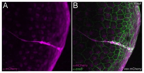
Labelling of Scaleforming odontoblasts by zns5 Lateral image of a 30dpf osx:mcherry transgenic immunostained for mCherry (magenta; A-B) and zns-5 (green; B), showing co-labelling of mineral forming odontoblasts on the scale |