
- Title
-
Chemically Induced Intestinal Damage Models in Zebrafish Larvae
- Authors
- Oehlers, S.H., Flores, M.V., Hall, C.J., Okuda, K.S., Sison, J.O., Crosier, K.E., and Crosier, P.S.
- Source
- Full text @ Zebrafish
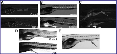
(A) Schematic outlining the area of intestine in 6 dpf larvae analyzed for enumeration of intestinal neutrophils. Images have been captured using low level background lighting with a GFP-long pass filter on a SMZ-1500 (Nikon) to demonstrate the feasibility of capturing and analyzing a single pseudo-overlay image. (B) Tg(mpx:EGFP)i114 6 dpf larvae. Top larva is phenotypically normal, bottom larva has ectopic GFP expression in intestinal epithelial cells as indicated by arrow. (C) Two DAF-FMDA stained 6 dpf larvae treated with 50 μg/mL TNBS to illustrate sites of inducible nitric oxide production. Constitutive staining is observed in the heart (easily identified by rhythmic beating), and is highlighted by the left arrow. Inducible staining is observed in the notochord (along the midline of the trunk) highlighted by the upper arrow, and in the cleithrum (in line with the developing gills) highlighted by the bottom arrow. (D) Alcian blue stained 6 dpf larvae. Top larva is phenotypically normal, bottom larva has been exposed to DSS and shows stereotypical accumulation of acidic mucins in the intestinal bulb, as indicated by arrow. (E) Neutral red stained 6 dpf larva. Arrow indicates location of endocytic mid-intestinal epithelium. |